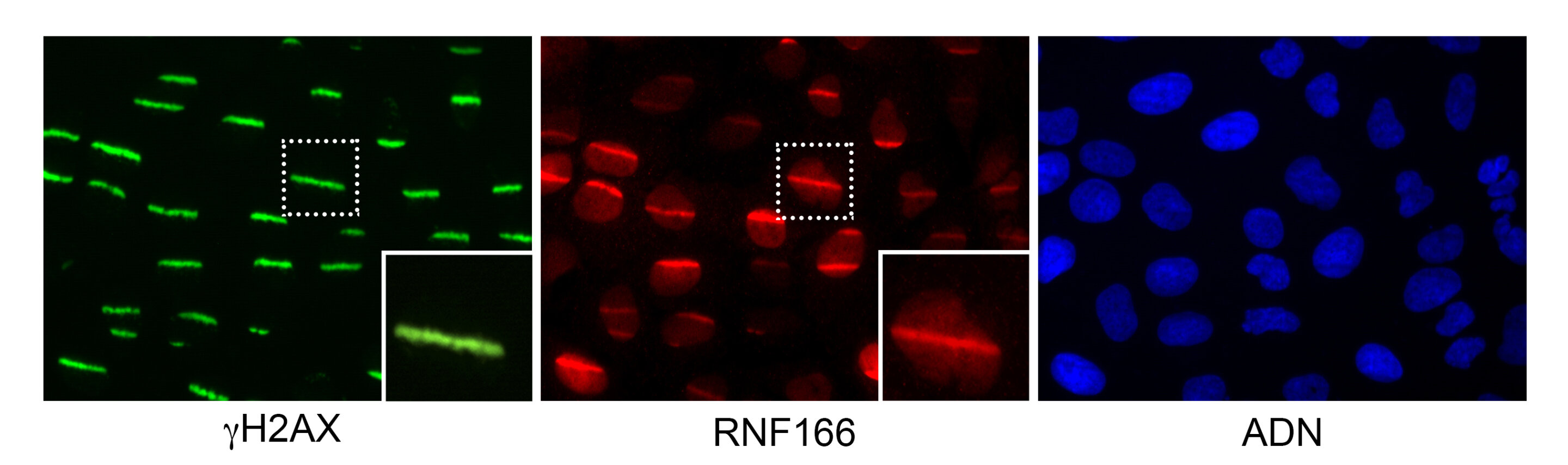
The human cells have their DNA damaged by a laser beam. In green, the classic gamma-H2AX repair protein bound to breaks generated by the laser beam; in red, the new protein discovered in this study bound to the DNA breaks. Credit: MGH.
The human body has trillions of cells and each one of them has at least 10,000 DNA problems a day. If the cells were unable to repair the injuries, they would be catastrophic, but there is a very delicate machinery that can detect and repair genetic damage. The National Cancer Research Centre (CNIO), led by Alejo Efeyan and Brbara Martnez, have been using machine learning to apply high-throughput microscopy. The results were designed in Boston and published in Cell Reports this week.
When there is a double-strand break, the cell's mechanism called the DNA damage response acts like a call to the emergency services. The alarm signals that are sent by the proteins rapidly bind damaged DNA and will be recognized by other proteins that are specialized in repairing the damage.
The goal of the drug is to kill the tumors by causing them to collapse and die. We can learn more about how cancer develops and how we can fight it by knowing how DNA lesions occur and how they are repaired. "Any new discovery in DNA repair will help develop better cancer therapies, whilst protecting our healthy cells."
The analysis of this process has never before been done with a degree of detail and precision thanks to the new methodology developed by the researchers. The inability to process and analyze the amount of data generated from images taken by the microscope is one of the limiting factors in tracking DNA repair.
High-throughput microscopy allows the acquisition of thousands of pictures of cells after they have been damaged. In the first phase, they introduced more than 300 different genes into the cells and tested whether they interfered with the repair of the DNA. Nine new genes have been discovered that are involved in DNA repair.
The video shows how one of the proteins discovered in this work targets DNA breaks. Credit: MGH.
The authors decided to look at the 300 proteins after they generated genetic damage. They adapted a classic DNA micro-irradiation technique to be used on a large scale format for the first time and analyze the behavior of the 300 proteins studied.
"We saw that many of the proteins that stuck to the damaged DNA moved away from the damaged DNA." The fact that they bind to or remove themselves from damaged DNA is a common feature of DNA repair proteins. Both phenomena are relevant.
One of the genes that was discovered is called PHF20. The authors showed that the recruitment of 53BP1, a key component in DNA repair, can be accomplished within seconds after damage. Cells without PHF20 are more sensitive to irradiation than normal cells, which indicates that they cannot repair their DNA properly.
These technologies can be used to study and manipulate DNA. Both platforms can be used to discover new genes or chemical compounds that affect DNA repair. We have used techniques that allow direct visualization of DNA repair in the evaluation of hundreds of proteins.
The work was funded by the Spanish Ministry of Science and Innovation, the Carlos III Institute of Health, the U.S. National Institutes of Health, the Marie Curie COFUND, and the Natural Sciences and Engineering Research Council of Canada.
Cell Reports has more information on the work of Raul Mostoslavsky. The full report can be found at www.cell.com/cell-reports.
Cell reports are in the journal.
Tools to visualize DNA repair were retrieved from thephys.org on December 28, 2021.
The document is copyrighted. Any fair dealing for the purpose of private study or research cannot be reproduced without written permission. The content is not intended to be used for anything other than information purposes.


