The crystal lattice has a large number of unit cells. In the elementary adiabatic picture, the motions of electrons in the crystal follow the motion of the atomic nuclei. The physical picture is valid for the core electrons of an atom, but fails for the valence electrons, which are shared by different atoms within its unit cell. The soft modes of phonons can change the electric properties of a crystal considerably. The properties of soft modes have been investigated for decades. Map atomic vibrations and charge motions at the same time is a must for a better understanding. This can be accomplished by using X-ray diffraction.
The researchers at the Max Born Institute in Berlin have been studying the motions of the electron and nuclear particles. The crystal is 500 times larger than the nuclear displacements, as reported in a recent publication in Physical Review Letters. The discovery of two related phenomena came from X-ray powder diffraction experiments on two prototypical crystals.
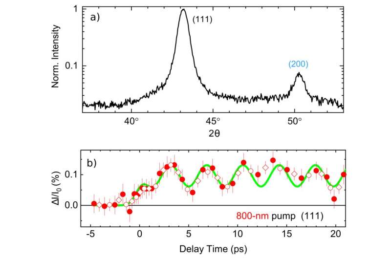
The team has implemented a method to take snapshots of the electron density in the unit cell of the crystal. It is possible to map nuclear positions and valence charge density on atomic length and time scales with the help of X-ray diffraction. In the experiments, an ultra short optical pulse causes atomic phonon motions in a powder sample, consisting of small crystallites. A snapshot of the momentary charge arrangement in the unit cell of the crystal is generated by the diffracted hard X-ray pulse. Changing the arrival time of the probe pulse allows for recording a pattern for each pump-probe delay, resulting in a movie of the promoted nuclear and electronic motions. The crystal remains in its electronic ground state because of the off-resonant impulsive Raman excitation.
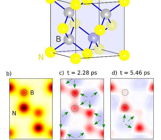


The figure shows the intensity of Bragg reflection from cBN. Transient electron density maps for different pump-probe delays show the relocation of valence electrons from the interstitial regions of the unit cell onto the atoms. The oscillations are caused by a coherent superposition of phonons.
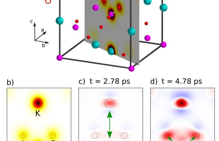
There are two pump-probe delays after coherent excitation of a soft mode. The swinging of electrons between the atoms in the ionic unit cell is caused by the motion of the nuclei. This behavior is very different from the predictions from literature and due to the longitudinal character of the nuclear motions. The electron density maps show a charge transfer between the K and P atoms and an electron relocation from the P to the O atoms.
The relocation of electronic charge occurs on the length scale of interatomic distances, whereas the underlying nuclear displacements occur on the sub-picometer. The crystal's energy content is reduced during the period when the phonon excitations are present. The findings serve as a benchmark for developing an adequate quantum description of soft modes and pave the way for future studies of a broad range of functional materials with ferroelectric properties.
More information: Shekhar Priyadarshi et al, Phonon-Induced Relocation of Valence Charge in Boron Nitride Observed by Ultrafast X-Ray Diffraction, Physical Review Letters (2022). DOI: 10.1103/PhysRevLett.128.136402 Citation: Persistent swinging of electrons between atomic sites in crystals (2022, April 1) retrieved 1 April 2022 from https://phys.org/news/2022-04-persistent-electrons-atomic-sites-crystals.html This document is subject to copyright. Apart from any fair dealing for the purpose of private study or research, no part may be reproduced without the written permission. The content is provided for information purposes only.All Rights Reserved © 2024


