The most notable feature of the disease is that it causes a wide range of symptoms beyond the respiratory distress. Symptoms vary wildly from person to person, from blood clot to loss of smell.
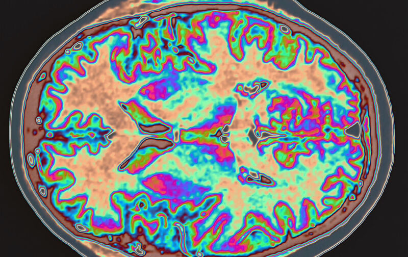
It will take years to figure out what the virus does inside the human body. We got some data this week from a study of the brains of COVID patients. The images were taken before and after the infections. The results show that some parts of the brain connected to the olfaction system may shrink in the wake of an infection, although the consequences are not clear.
This study uses the UK's Biobank. Users of the UK's National Health Service can link their medical records to their genetic profiles in order to provide medical researchers with a resource of large, population-level studies of risk. A research team based in the UK combed the Biobank for people who had had brain scans prior to the disease.
There are many reasons to focus on brains. Loss of smell is the most obvious. We don't currently understand the biological reason for this, but it appears that the nerve cells in the nose are affected. There is only one nerve bundle that connects to the areas of the brain that process smell signals. The nerve bundle could transmit the virus to the brain.
The brain can be altered by an infection. Damage due to the blood clot seen in some patients can be caused by setting off an inflammatory response in the brain. There are reasons to think that something is going on, as there have been reports of a poorly defined brain fog that some people experience. A number of small studies have shown the damage to the brain after an infection.
AdvertisementThe researchers wanted to get a better picture of the brain, so they got over 400 people who had received a brain Scan prior to the outbreak. The patients were put with people who had not tested positive for the virus to get brain scans and serve as controls.
The researchers looked for changes in specific structures using a software package that was able to compare two brain images. They did two separate analyses. One was hypothesis driven, and it was limited to all the structures that are known to be connected to the olfactory nerves in the nose. The second search was for any brain structures that differed from the before and after scans.
The differences between the two time points in patients who had been infections were small. It would take about five years for the brain to shrink due to the natural decline that occurs with aging. The difference appeared to be due to changes in the brain's gray matter, rather than the white matter used to establish connections between cells.
Most of the changes were seen in people who had been bitten. This is not a case where only a few people experienced large-scale changes that threw the numbers off. The data was reanalyzed when 15 patients who had required hospitalization were removed. The changes do not seem to need a severe case of COVID-19.
The hypothesis-drive analysis shows a strong correlation between the changes identified in the general analysis and the olfactory system.