John Hewitt is a writer for Phys.org.
Credit: Wikimedia Commons
Many human diseases are caused by jumping genes. Keeping them repressed by the immune system is a full-time jump for cells.
Last week, we looked at the activation of the Line-1 retrotransposons in a host of neurodegenerative conditions. Human retroviruses have a long terminal repeat sequence at the beginning and end of their genes.
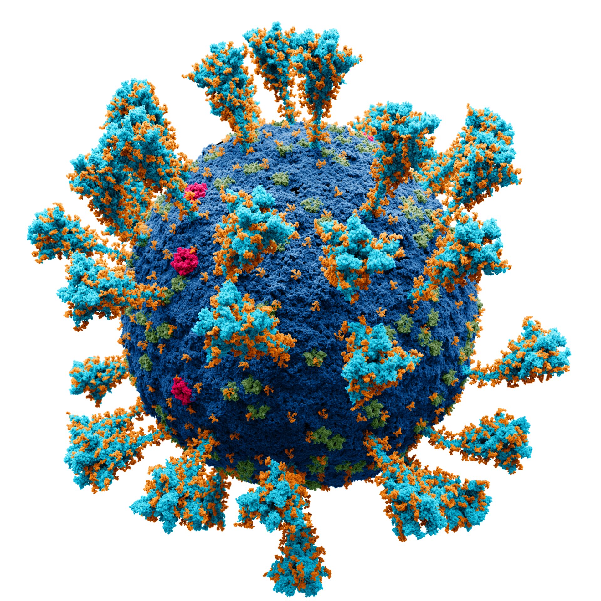
On Tuesday, a real zinger was dropped onto the medRxiv preprint server that could potentially explain many of the commonly observed pathogenic features of SARS-CoV-2. The authors provide solid evidence that the envelope ENV is activated in blood cells by the SARS-CoV-2 spike, which is in turn responsible for many pathological features of the disease. Many retroviruses in the group use a tryptophan in the primer binding site, which is why HERV-W is named. The shape of the letter W reminded the naming committee of the ring structure of atoms in the side chain of tryptophan.
There is a correlation between the expression of HERV-W ENV in T lymphocytes and the severity of respiratory distress in patients with coronaviruses. The mechanisms involved were not clear. The real culprit in HERV-W activation has been found. The cultured peripheral blood mononuclear cells (PBMCs) from patients with the disease were added to the database. The upregulation of the ENV protein from both HERV-W and HERV-K was immediately found. Only the HERV-WRNAs resulted in the expression of ENV.
There are slight modifications that make the mRNA less immunogenic and the spike protein more immunogenic. Adding two strategic prolines to the code is one way this has been done. More research is needed to fully understand the fusogenic potential of spike proteins. Some vaccine manufacturers have eliminated the furin cleavage site from their constructs in order to reduce the risk of residual fusion. An anonymous researcher on social media pointed out a few of these observations to me.
The studies show that not all COVID patients had HERV-W ENV activation. This finding likely reflects an underlying genetic susceptibility among the infections that needs to be defined and taken into account, particularly if HERV-W is going to be used as a general marker for disease severity, or as a therapeutic target for a humanized monoclonal antibody therapy. The HERV-W form that was found in multiplesclerosis is a potential druggable option.
Which HERV-W, exactly? The HERV-W remnants make up over 1 percent of our genomes. There are at least 13 HERV-W genes in the human genome. There is an open reading frame for a complete HERV-W ENV protein in one of these. This is a well-known and well-defined part of normal placental development. To complicate things, it seems thatMS has many different possible origins. This week, researchers revealed that an infection with the Epstein-Barr virus is an important upstream, or downstream, or perhaps independent,trigger for multiplesclerosis.
There are other retroviral games in town. Researchers recently discovered that a retroviruses-likeProtein known as PEG10 directly bind to and produce its own mRNA in capsids. This behavior is similar to that of the ARC1 retroviral protein, which is believed to be critical in the formation of memory at synaptic sites. Researchers are well on their way to pseudotyping these virus-like particles with fusogens to create an endogenous vector for delivering functional mRNA cargos as a gene therapy. Caution is needed in these affairs.
HERV-W ENV was mostly found in cells from small blood vessels in the heart tissue samples. The HERV-W ENV positive cells were confirmed with CD31 staining. Significant HERV-W ENV was found in the central nervous system and in the blood clot in patients, even though there was no evidence of the disease in those tissues. The authors note that the HERV-W ENV expression in human lymphoid cells is caused by the SARS-CoV-2. This suggests other routes for the virus. One recent clue to other candidate mechanisms might be the expression of ASGR1, which is highly expressed in liver cells.
It is important to find out how HERVs are activated. In light of the known penchant for transposable elements to be activated by and further integrate into sites of active DNA repair, it may be worth revisiting earlier studies that purported to show that reverse transcribed SARS-CoV-2 RNA could integrate into the genome of cultured human cells. The target site duplications were found to be consistent with the LINE1 retrotransposon-mediated, target-primed reverse transcription and retroposition mechanism.
Benjamin Charvet and his co-workers have shown that the human retroviruses type W envelope are present in the blood and tissues of COVID-19 patients. There is a book titled "10101/2022.01.18.21266111".
The Science X Network is in the year 2022.
There is a report about a spike in the number of human retroviruses in blood cells.
The document is copyrighted. Any fair dealing for the purpose of private study or research cannot be reproduced without written permission. The information provided is for educational purposes.
All Rights Reserved © 2024


