The veterinarians at Chicago's Shedd Aquarium have pioneered a lot of unusual procedures to diagnosis and treat the animals in their care.
We reported on the case of the missing chloroquine at the Shedd Aquarium. The antiparasitic drug is added to the water for new animals, but it was mysteriously disappearing. The post included a line about how the aquarium vets had the lowdown on how to give an electric eel an MRI.
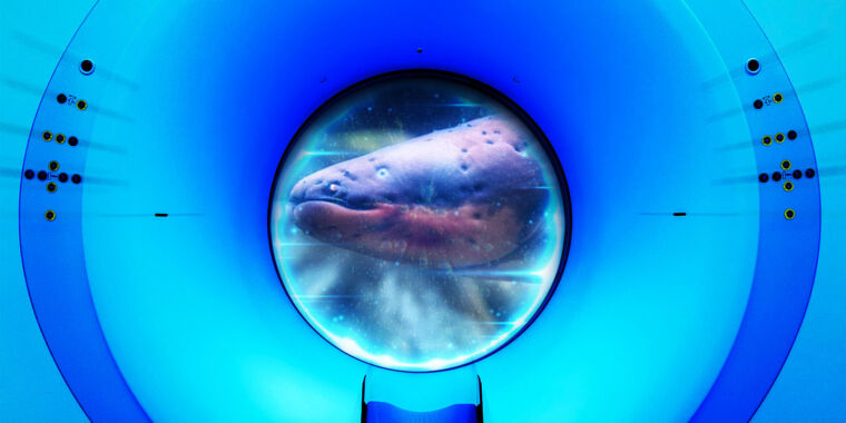
We received several queries about how this feat might be accomplished, after we heard about that bit. You asked. We wanted to know more. The Center for Animal Health and Welfare at the aquarium has a state-of-the-art animal hospital that is used to monitor the health of all the animals in the exhibits. Dr. Van Bonn and Dr. Karisa Tang were happy to help.
Van Bonn says the veterinary team at the aquarium is more of a family practice than a specialist. Comparative medicine is practiced by necessity since there isn't a lot of precedent in the literature for many of the animals in their care.
Electric eels are technically knifefish. Three pairs of abdominal organs are located symmetrically on both sides of the eel and are used to produce electric discharges. The brain opens ion channels and reverses the polarity when it sends a signal. A battery with stacked plates is similar to the difference in electric potential generating a current.
It's that ability that makes it difficult to give the creatures an MRI. The creatures can vary the degree of voltage in their electrical discharges, using lower voltages than a car battery, to sense their environment, navigate, and hunt. They have tiny eyes because they live in murky waters where there is not much light or visibility. The eels use higher voltages to stun and kill their prey.
Advertisement
Van Bonn wondered if he and his staff would need to worry about the higher voltage discharges since the aquarium eels are in captivity. In managed care, poison dart Frogs don't produce their trademark poison because they don't need the defense mechanism. He put a voltmeter in the water. It had a low potential. When he poked the animal, the reading on the voltmeter went up. The eels need to be handled with care.
Electric eels are capable of generating high voltages to stun and kill prey.
An electric eel discharge could cause an artifact on the resulting image that renders it useless. Nobody wants the handler to die. Figuring out a way to anesthetize the eel is the first step. The handler must wear gloves and footwear for the process to stop.
The vets at the aquarium usually put a powder in the water to anesthetize fish, unlike humans who breathe it in via a gas. The team uses the same method as they do for other fish, because eels breathe air but also breathe through the water with their gills. Correct dosages must be determined through trial and error, and the team should be aware of any discharges if the animal isn't completely asleep. Any nonelectric fish or tubular animal can be anesthetized and the magnetic resonance image can proceed.
The vets have to give little puffs of oxygen over the oral cavity since an anesthetized eel can theoretically drown. The air travels through a trachea to the lungs so we can breathe, but electric eels have wrinkled tissue on the inside of their mouths. "You just have to breathe in the tissue with the puff oxygen."
The unhappy trio of O'Donoghue.
Is it possible to perform knee surgery on a bullFrog? We can do that.
You're going to get a lot of good stories when you treat weird, exotic animals. Van Bonn said that they learn something new every time they work with an animal. Whenever the Shedd Aquarium acquires new animals, they are placed in theQuarantine habitat to prevent them from introducing any outside pathogens into the aquarium's carefully controlled environment.
Advertisement
One day, an aquarist brought Van Bonn a bullFrog that had hurt its leg while wrestling with another male of the species. The injury was similar to a blown knee, according to Van Bonn. This injury occurs when there are tears in the knee joints. He said that the leg is flopping out to the side. It's a serious injury that's known as the unhappy triad of O'Donoghue.
Human knee surgery is common and documented in the medical literature. Van Bonn didn't know if the frog had a knee injury. Nobody writes about the surgical part of a bullFrog's leg.
He found the diagram in a 19th-century German book. Van Bonn had to make his own artificial froggy ligaments to repair his knee. BullFrogs require six times more anesthesia to knock them out than fish, for some strange reason, despite being sensitive to environmental contaminants.
Beluga whales can't be transported to the second floor of the hospital via elevator. Their habitat has a platform that can be raised.
What if the animal needs treatment that is larger? The animal hospital is located on the second floor of the Shedd building, and a beluga can't be transported in the elevator. The behind-the-scenes care area is located in every habitat in the aquarium. There is a special platform that can be raised if needed at the Oceanarium, where the belugas live.
Taking blood samples can be done in situ with the help of the staff. Commercial horse endoscopes work just fine, even though they aren't specifically designed for beluga whales.
"There are years and years of experience and training behind all these one-off stories about how do you reconstruct a bullfrog's knee, or how do you build a nebulizer for an anaconda, or what level of pain medication will help a shark," said Tang. The team members at the Shedd Aquarium are dedicated to publishing scientific studies about their work to help build up the literature and spread what they've learned to other zoological and aquatic veterinarians around the world.


