The human lung epithelium cell was damaged by the viral infections. There are jacked cellular organelles. Venera Weinhardt is aCOS.
The interior structure of the cells that are attacked by the coronaviruses change. The changes at the level of individual cell components can provide information on how diseases develop. Powerful techniques are needed to visualize them, but they are very data- and time- intensive. A research team under the direction of Dr. Venera Weinhardt at the Centre for Organismal Studies (COS) at the University of Heidelberg recently improved a special X-ray process to deliver high-resolution three-dimensional images of entire.
The Lawrence Berkeley National Laboratory in Berkeley, USA, has a post-doc named Venera Weinhardt who says that scanning electron microscopes are preferred in cell images. This technology takes a week to do. It is difficult to analyze and interpret the huge amount of data it creates. We get usable results in five to ten minutes using soft X-ray tomography. Prof. Dr. Bartenschlager is working with Dr. Weinhardt to study cellular changes associated with viral infections. The scientist says that in tissue only a small amount of the cells are infectious. Only those cells give information about the changes that occur. It is not possible to look for these cells with a scanning electron microscope.
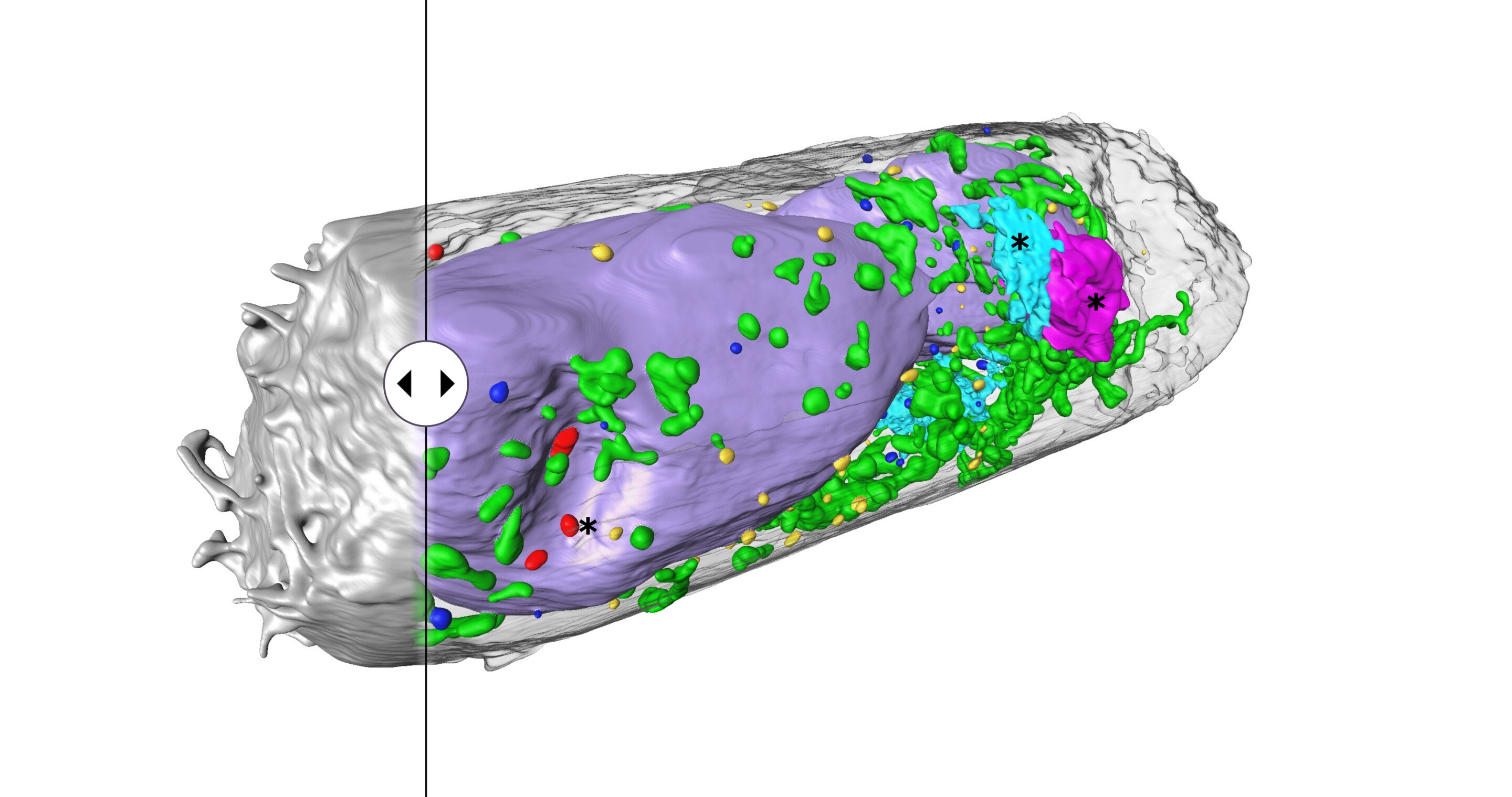
The procedure known as soft X-ray tomography (SXT) has already been used to detect single virus particles that are different from each other. The researchers used the technology to study cell cultures that had been exposed to the disease. They were able to see complete cells and their structure in five to ten minutes. The researchers were able to detect clusters of particles on cell surfaces as well as identify changes in the cell's interior. The structures may enable the spread of the virus.
The team's success was largely due to the technology that allowed them to study fixed cells. Flat lattice structures are usually used as holders in soft X-ray tomography. The thickness of the samples can change when they are tilted. The flat shape of the holder prevents the cells from being scanned at all angles. The sample can adhere to the lattice or spread out, but it requires multiple tomograms to see the entire cell. To get around the problem, we switched to cylindrical thin-wall glass capillaries. The researcher says that the samples can be scanned from all angles. The team is working on a soft X-ray microscope, sample preparation techniques, and automated analysis of 3-D image data.
The results of the research were published.
More information is provided by the authors of " Using soft X-ray tomography for rapid whole-cell quantitative imaging of SARS-CoV-2 cells." The article is titled "Creme.2021.100117."
The news about visualising cell structures in three dimensions was retrieved fromphys.org on December 6.
The document is copyrighted. Any fair dealing for the purpose of private study or research cannot be reproduced without written permission. The content is not intended to be used for anything other than information purposes.


