Scientists in the United States have created a new drug that stimulates cell regeneration and reverses paralysis in spinal injury mice. This allows them to walk again in four weeks.
The Northwestern University researchers behind the research were published in Science on Thursday. They hope to contact the Food and Drug Administration (FDA), as soon as possible, to suggest human trials.
Samuel Stupp from Northwestern, who was the lead of the research, said that the goal of the study was to create a translatable therapy to help people avoid paralysis after major traumas or diseases.
Paralysis treatment is an ongoing goal of medicine. Other cutting-edge research includes gene therapy, which tells the body to make certain proteins in order to assist nerve repair and gene therapy.
Stupp's team used nanofibers to imitate the extracellular matrix, a network of molecules that surrounds tissue and is responsible for supporting the cells.
Each fiber is approximately 10,000 times thinner than a human hair and contains hundreds of thousands bioactive molecules called "peptides" that transmit signals to stimulate nerve regeneration.
After an incision in the spines of laboratory mice, the therapy was administered as a gel to the tissue around their spinal cords.
Illustration of biomolecules (green & orange) communicating with cells to repair damaged spine cord. (Mark Seniw)
Because of the delays that can occur when people receive severe spinal injuries from gunshots, car accidents, and other causes, the team decided to wait.
The treatment was effective for four weeks. After the procedure, the mice were able to walk almost the same as before the injury. The treatment was not effective for those who were left untreated.
The therapy was then applied to cells, and the mice were put to sleep.
Scar tissue, which can be used to block regeneration, was significantly reduced by the regeneration of the axons, or severed extensions of neuronal neurons.
Furthermore, myelin, which is an insulating layer of the axons that transmit electric signals, had been reformed. Blood vessels that supply nutrients to injured cells were also formed. More motor neurons survived.
"Dancing" molecules
The team discovered that a specific mutation in the molecules could increase their collective motion and enhance their efficacy. This was a key discovery.
This is because neurons have receptors that are in constant motion. Stupp explained that increasing the motion of therapeutic molecules in nanofibers can help them connect more effectively to their moving targets.
Researchers actually tested two versions, one with and one without the mutation. They found that mice who received the modified treatment had greater function.
(Samuel I. Stupp Laboratory/Northwestern University)
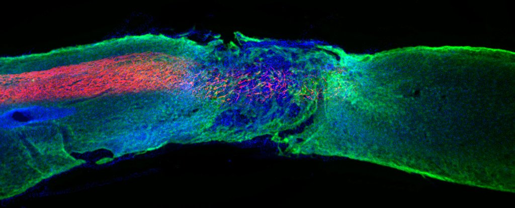
Above: 12 weeks after an injury, red blood vessels are growing through the spinal cord cells (blue).
Scientists created the gel, which is unique in its type. However, it could be the start of a new generation of medicines called "supramolecular drug" because the therapy is made up of multiple molecules, rather than one.
The team claims that it is safe as the materials can be biodegraded in a matter weeks, and then become nutrients for the cells.
Stupp stated that he plans to quickly move to human studies without further animal testing.
It is because of the similar nervous systems across mammal species. He said that "there is nothing available to help patients with spinal cord injuries, and this is an enormous human problem."
Official statistics show that nearly 300,000 Americans have a spinal injury. Their life expectancy is much shorter than that of people with no spinal injury and has not increased since the 1980s.
"The problem will be how FDA will view these therapies since they are completely new," said Stupp.
Caption: Spinal cord (green, blue) with regenerative Axons (red), flowing towards central damage.
(c) Agence France Presse
