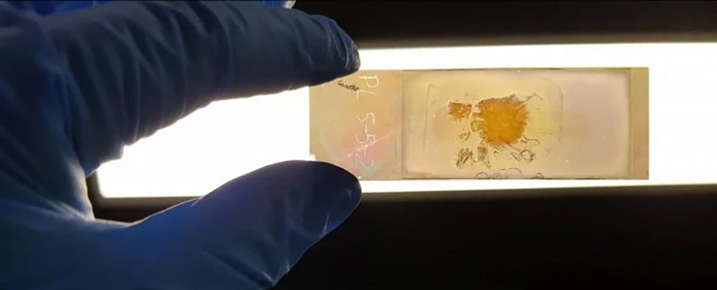
The NanoMslide is a five-year-old invention. It can work with a standard microscope to identify cancer cells in distinct colors. Experts are able to detect signs of the disease more quickly.
This new technology is based upon the physics and oscillations of charged particles such as electrons, in the physics known as plasmons. Thin silver layers provide entire fields of free-roaming, electron-rich areas that, when stimulated with light, align in a particular way.
By poking tiny, nanosized holes in the silver, you can manipulate the light's structures. As the light passes through tissue, certain colors are selected. This creates a spectrum that is dependent on subtle differences in the structure of the sample and, importantly, its electron configurations.
The microscope slide is now a sensor and can reveal the cell composition through a color-coded lighting show. It will allow for quicker diagnosis of disease, and without the need to apply stains or risk damaging samples.
Under the microscope, cancerous cells are highlighted (La Trobe University)
Belinda Parker, a biochemist from La Trobe University in Australia, says that she was thrilled to see a piece of tissue under the microscope using the NanoMslide for the first time.
"For the first-time, I saw cancer cells popping up right in front of me for the first time." They had a distinct color and were easy to differentiate from surrounding cells.
Parker and her colleagues described the change as a leap from black and white televisions to colors. It can be difficult to find cancer cells using the existing methods.
To create their slides, the team used advanced fabrication techniques that were not yet available. The technology should be useful in many medical and non-medical settings once it is scaled up.
Nanotech slides are much faster than other techniques and can produce results in as little time as 10 minutes. It's extremely helpful in time-critical situations to decide which parts of a tumour should be removed during surgery.
Brian Abbey, a physicist/chemist from La Trobe University, says that current approaches to tissue imaging often depend on labeling cells or staining them to make them visible under the microscope.
"Even with labeling and staining, it can be difficult for pathologists to identify cancer cells. There is a risk of some samples being misdiagnosed especially in the early stages.
The NanoMslide is extremely effective, but the researchers aren't sure exactly what makes the colors different. They suggest that it could be proteins or cell skeletons.
The researchers have already completed a study that shows how nanotechnology can improve the process of identifying early-stage breast carcinoma from benign lesions. They used both human tissue and a mouse model.
The NanoMslide team is confident that it can be modified to detect different types of cancers, and even other diseases. The tech's creators are working on commercializing it.
Abbey states, "Our vision is for our technology to help the diagnosis of a variety of other cancers through analyzing all kinds of tissue sections as well as use to plant biology and agriculture."
Nature published the research.


