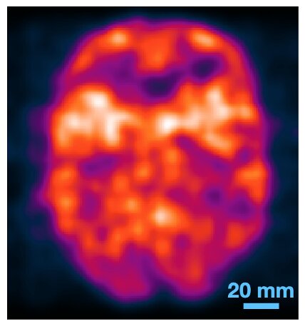
Ultrafast photon detectors are now capable of processing data from positron emission and X-ray scans quickly without the need to use tomography to reconstruct images. This image shows a brain model (or brainphantom) that was scanned using positron emission. Credit: Simon Cherry, UC Davis
Japan and the U.S. have created the first cross-sectional experimental medical image. This is a mathematical process that reconstructs images in CT and PET scans. This work was published in Nature Photonics on Oct. 14. It could make medical imaging more affordable, simpler, and more accurate.
Simon Cherry, professor of radiology and biomedical engineering at the University of California, Davis, and senior author of the paper, stated that the breakthrough was possible because of the development of ultrafast photon detectors.
Cherry stated, "We are literally imaging at the speed light, which is something that's something of a holy ground in our field."
Sun Il Kwon was the project scientist at the UC Davis Department of Biomed Engineering. Ryosuke Oka at Hamamatsu Photonics in Japan was also involved in experimental work. This is where the new photon detection technology was created. Researchers led by Professor Yoichi Takamawa at the University of Fukui and Professor Tomoyuki Hasegawa from Kitasato University were also collaborators.
Tomography is a mathematical process that reconstructs cross-sectional images using data from imaging that uses X or gamma radiations. PET scans use radioactive molecules that have been tagged to be injected into the body and then taken up by tissues and organs. Fluorine-18 is an unstable radioactive isotope that emits positrons when it decays.
Ultrafast photon detection
When one of these positrons meets an electron in the human body, they annihilate and simultaneously emit two annihilation photosns. The theory is that these photons can be tracked to create an image of tissues tagged with itotopes. Researchers were not able to do this without tomographic reconstruction. This was because detectors were too slow for precise detection of arrival times and could not pinpoint their locations based on time differences.
Cherenkov photons are produced when annihilation photos hit the detector. Cherry and his colleagues discovered a way to detect Cherenkov photons at an average timing precision 32 picoseconds. They were able to determine the origin of the annihilation photos with a spatial precision measuring 4.8 millimeters. The team was able to create cross-sectional images of radioactive isotopes directly from the annihilation photos without the need for tomography.
The researchers present a variety of tests that they performed using their new technique in their paper. One test was on an object that mimics the human mind. The researchers feel confident that the procedure can be scaled to clinical diagnostics levels and can produce higher quality images with a lower radiation dose. This method allows for faster creation of images, possibly even during the PET scan.
PET scans can be expensive and limited technically. The full information in the travel times of the annihilation photosns is not captured by the current clinical scanners. The new technology could allow for accurate, inexpensive scans of the human body with radioactive isotopes.
Eric Berg, University of California Davis; Fumio Hamshimoto and Tomohide Omura, Hamamatsu Photonics, Kyohei Nakajima, and Izumi Okawa, University of Fukui are additional coauthors.
More information: Sun Il Kwon et al, Ultrafast timing enables reconstruction-free positron emission imaging, Nature Photonics (2021). Journal information: Nature Photonics Sun Il Kwon et al, Ultrafast timing enables reconstruction-free positron emission imaging,(2021). DOI: 10.1038/s41566-021-00871-2


