There is always more to discover and learn. This holds true whether you zoom in to tiny organisms or out into the vast reaches of our Universe. Science is a process where you ask more questions than you answer.
Researchers have now taken their powerful microscopes to the skin of bacteria and are now able to see the inner workings of the membrane, providing more information than ever.
The outer membranes of Gram-negative bacteria such as Escherichia Coli are used to protect their cells from the bacterial world. These membranes contain a great variety of tools, including outer-membrane protein and toxic substances like lipopolysaccharides.
Once you have this information, there are still questions about how all of this amazingness fits together. How many'studs to membrane are there in the entire bacterium? This is where new research steps in.
"The outer membrane acts as a strong barrier against antibiotics, and is an important factor in making infections resistant to medical treatment. Bart Hoogenboom, a University College London biophysicist, said that it is still not clear how the barrier is constructed.
"By studying living bacteria at the molecular and cellular levels, we can see how membrane protein networks span the entire bacteria's surface, leaving little gaps for areas that lack protein."
For this purpose, the researchers used an atomic force microscope. This microscope continuously pokes at the membrane's surface to see what it looks like. It's almost like reading braille with lasers instead on your fingertips.
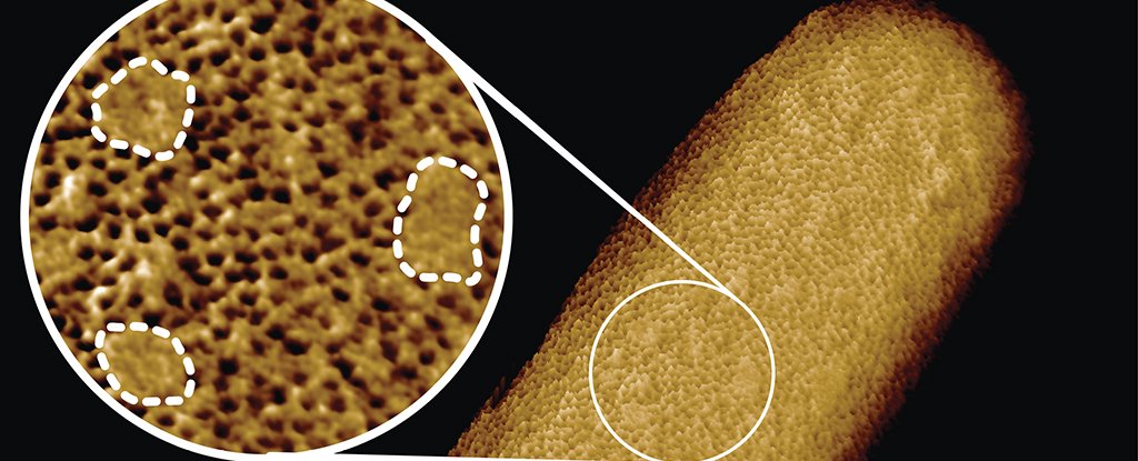
The team produced an amazing image of the bacterial membrane, which they called the sharpest ever taken of living bacteria. It shows the density of the outer-membrane protein across the surface.
The outer membrane is visible using atomic force microscopy. (Benn et al., PNAS, 2021)
You can see that there are many tiny holes on the surface, as you can see from the above image. These holes are the beta barrel proteins (or porins) that create tunnels in bacteria's membrane for molecules to pass through. The lipopolysaccharides, which can expand with cell growth, are located on the small portions of the smooth surface.
"We conclude that the outer membrane is a mosaic of phase-separated lipopolysaccharide-rich and outer-membrane protein-rich regions, the maintenance of which is essential to the integrity of the membrane and hence to the lifestyle of a gram-negative bacterium," the team writes in the new paper.
Scientists don't just stare at E.coli with giant microscopes for fun. This level of detail is necessary for many reasons.
"The classic picture of the outer membrane of bacterial bacteria shows proteins distributed in a disordered fashion, well-mixed together with other building blocks. The images show that this is not true. However, lipid patches are separated from protein-rich networks in a manner similar to oil separating form water. In some cases, chinks are formed in the armor of bacteria.
"This new approach to the outer membrane allows us to now explore if and how this order affects membrane function, integrity, and resistance against antibiotics."
This team will investigate how to use the new knowledge to overcome antimicrobial resistance in gram negative bacteria like E. coli.
As with the black holes and stars in far-flung galaxies there is always more to discover.
The research was published in PNAS.


