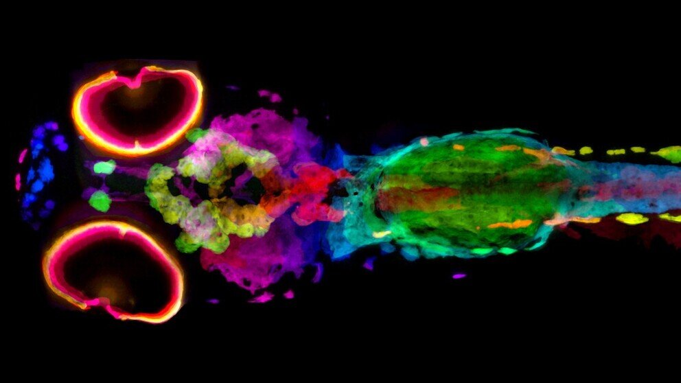
A 5-day-old, silver-stained zebrafish is used to visualize melanin using X-ray histotomography. Based on the amount of melanin present in the cells, the colors were determined. Credit: Keith Cheng Lab at Penn State College of Medicine
Scientists and doctors require quantitative, three-dimensional information to understand the biology of skin pigmentation and other related diseases like albinism and melanoma. Researchers at Penn State College of Medicine have created a new method that allows scientists to see every melanin pigment-containing cell in 3D in whole Zebrafish.
Because melanin blocks light, it is difficult to study. Researchers turned to Xray imaging for this purpose, as it can penetrate optically opaque material like melanin.
A team of distinguished pathologists led by Dr. Keith Cheng (professor of pathology and pharmacology, biochemistry, and molecular biology), developed "Xray histotomography", a cell-based form of CT imaging. This was a way to examine the 3D structure of cells and tissues in biological samples with unprecedented clarity and resolution. Spencer Katz, an MD/Ph.D. Cheng's medical scientist training program student has modified the micro-CT technique in order to investigate melanin. This pigment is being studied in human skin color research and melanoma research in whole Zebrafish.
Melanin, a brown-to-black pigment that gives the Zebrafish their distinctive stripes, and humans their dark skin and hair. Cheng and his laboratory discovered a key gene that controls the evolution of light skin color in humans more than 15 years ago by studying a zebrafish mutant, called golden. This particular zebrafish line has lighter stripes. This discovery proved the importance of zebrafish models in studying human biology and disease, such as albinism or melanoma.
Like human CT, micro-CT uses a series X-rays from slightly different angles to reconstruct 3D representations. The micro-CT samples are smaller, and the resolution Cheng's group has developed is 2000 times higher. Katz used silver to stain melanin. This allowed researchers to determine 3D location of melanin and its density from scans of whole Zebrafish.
The Cheng Lab collaborated with Dilworth Parkinson, at the Advanced Light Source at Lawrence Berkeley National Labs, Berkeley, California. He directs a microCT resource that Cheng can use for X-ray histotomography. The new X-ray detector system at the lab was created to provide unprecedented resolutions for samples as large as whole zebrafish and human biopsies. The team tested zebrafish with normal and altered pigmentation (including golden).
Researchers were able to see every melanin-containing cell in fish and map their locations in 3D. They were also able to take quantitative melanin measurements, which allowed them to compare melanin levels between normal and mutant fish. The results of the study were published in eLife.
This research laid the foundation for further research into melanin-containing cancers or melanomas. These are usually graded according to the extent of tumor cell invasion. Cheng, a Penn State Cancer Institute researcher, said that a variety of zebrafish models for melanoma can be used to study the new technique. Katz and Cheng stated that silver can be used to stain human melanomas and images of them in the same manner can be taken. This will allow for more detailed analysis of the tumor cells and their arrangement in the tumors. For example, scientists will be able to count tumor cells with different characteristics and study invasion more thoroughly, which is a key characteristic of cancer. This will help doctors make better prognostications and treatment decisions.
The Cheng Lab will continue to improve the application of histotomography by developing new optical and staining methods. This study provides proof-of-principle that whole-body 3D computational analysis can be done on tissues and organisms using micro-CT. This may enable a more comprehensive understanding of gene function.
This research was also done by Maksim Yakovlev and Daniel Vanselow. Yifu Daing, Yifu, Alex Lin, Victor Canfield, Khai Chung Ang, Yifu, Ding, Yifu, Ding, Alex Lin, Victor Canfield, and Yifu. Yuxin Wang, Mobile Imaging Innovations Inc. designed and built the new lens system. The authors declare no competing interests.
Continue reading
More information: Spencer R. Katz et., Whole-organism quantitative characterization of zebrafish melanin by silver deposit micro-CT, eLife (2021). Information from eLife Spencer R. Katz et. al. Whole-organism 3-D quantitative characterization zebrafish melanin using silver deposition microCT, (2021). DOI: 10.7554/eLife.68920


