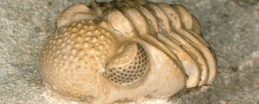
Half a century ago, an amateur paleontologist first examined a fossilized trilobite. This discovery has given researchers a new way to see the world in a literal sense.
The second look at X-rays of the ancient arthropod taken in the 1970s has revealed a structure that is unique to any eye ever seen on an animal.
Wilhelm Strmer was the head of Siemens radiology department and knew a lot about using Xrays to uncover hidden secrets. This was particularly true when it comes to fossil study, which Strmer fueled with the creation of a minibus equipped with X-ray equipment that could be taken to sites for paleontology.
Despite his radiology expertise, Strmer was not a paleontologist. Therefore, few people took Strmer's claim that he had discovered optic nerves in a Phacops geesops fossil dating back 390-million years old, despite his radiology expertise.
Brigitte Schoenemann, University of Cologne paleontologist, says that at that time it was agreed that only bones and teeth could be seen in fossils. However, they did not include the soft parts like intestines and nerves.
A group of 'fibers,' which looked very much like photoreceptor cells, was found in addition to the nerves. They were only 25 times longer than their diameter in this instance, making them impossible to be considered light-gathering structures.
Since then, a lot has happened. Paleontologists today are more comfortable accepting the fact that fossilized material can be traced back to soft tissue structures. In the compound eyes of aquatic arthropods, super-long ommatidia were also discovered.
Schoenemann and her coworkers re-examined Strmer's original images to get a better look. They confirmed that the filaments he saw were optic nerve fibers, after double-checking it with modern CT technology.
Researchers were attracted to the foam-like nest of fibres that was connected to it. What appeared like two compound eyes, actually contained hundreds of them, divided into left and right clusters.
Schoenemann says that each eye contained approximately 200 lenses, with a maximum size of one millimeter.
"Under each one of these lenses are at least six facets, each of which together make up a small compound eyes. We now have around 200 compound eyes, one under each lens.
Trilobites dominated the oceans for hundreds and millions of years. They adapted to fill many aquatic niches with a wide range of bizarre and wonderful body plans.
Their vision system was an astonishingly complex invention. Modern eyes are simple, but their vision system was more complex than modern. It allowed them to hunt or hide and detect subtle changes in brightness or movement.
Although their eye anatomy was complex, most would recognize the most common ones. This is a pattern of lenses that works together to transform diffuse light into a highly pixelated map.
Today's insects and other arthropods still rely on compound eyes to great effect. This pixelated view is not as sharp as it could be, but the simplicity and adaptability of this compound eye makes up for that.
Yet, paleontologists have been baffled by the extraordinary diversity of trilobite eye colors found on Phacopina suborder members.
A schizochroal lens is a lens that sits close to its neighbor. This creates an empty space which could be used for catching more light.
Now we know that what appears to be one lens is actually two compound eyes within two 'hypereyes'.
Although it doesn't explain how these eyes evolved it does alter the questions we should ask about this unique arthropod.
Instead of pondering how much space is wasted between each lens, biologists now have the opportunity to speculate about the many benefits hundreds of tiny eyes can provide in adapting to low light or responding to sudden changes in light conditions across a larger area.
Schoenemann says that it is possible for the different components of an eye to perform different functions. This would allow, for example, contrast enhancement and the perception of different colours.
Strmer must have known there was something to be seen in the eyes. He had drawn an arrow with red pen, pointing directly at half-dozen of the facets below one of the lenses.
Unfortunately, the radiologist died in the 1980s, well before his discovery received the recognition it deserved.
Strmer, like the trilobite was clearly a visionary in his own right before his time.
This research was published by Scientific Reports.
