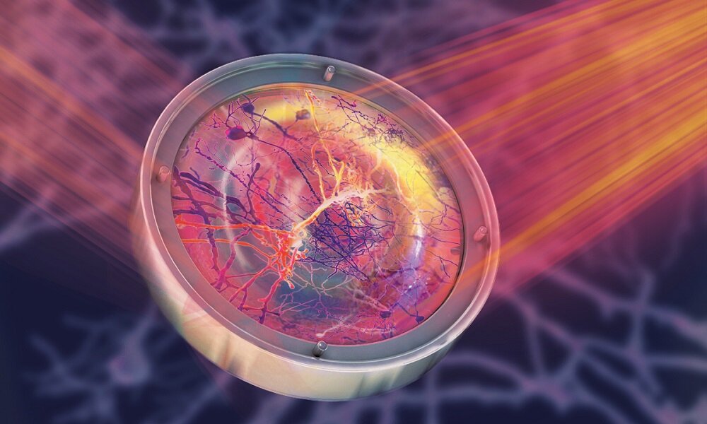
Microscopy uses a deformable mirror to focus light on live tissue. They would normaly distort its propagation. This mirror allows us to see the neurons deep within the brain in clear, sharp images. Credit: Isabel Romero Calvo/EMBL
The Prevedel Group at EMBL has developed a groundbreaking technique that allows neuroscientists access to live neurons deep within the brain, or any other cells hidden in opaque tissue. This technique uses two state-of the-art microscopy techniques, three-photon microscope and adaptive optics. Nature Methods published the paper describing this breakthrough on September 30, 2021.
It was difficult for neuroscientists, prior to the invention of the new technique to see astrocytes creating calcium waves in deep layers in the cortex. They also had to visualize any other neural cells within the hippocampus. This is a deep part of the brain responsible for navigation and spatial memory. This phenomenon is common in all living mammals' brains. Lina Streich, from the Prevedel Group, and her collaborators developed a new technique that allowed them to capture the intricate details of these flexible cells at an unprecedented high resolution. Researchers from India, China, India, France, USA, India and Austria were part of the international team.
Brain tissues are mostly observed in neurosciences in small models or ex vivo specimens that must be cut up for observation. Both of these conditions are non-physiological. Robert Prevedel stated that normal brain cell activity occurs only in living animals. However, the "mouse mind" is highly scattering. Because light interacts with the cells, it is difficult to focus in these brains. This makes it difficult to focus on the small structures deep within the brain using traditional techniques.
Scientists can now see deeper into tissue thanks to Streich, who was a former PhD student in our lab and worked for over four years to solve this problem.
Prevedel, who is a trained physicist, explained that traditional fluorescence brain microscopy techniques absorb two photons each time and can ensure that the radiation's excitement is contained to a small volume. "But scattering can make photons lose more often the farther they travel." This can be overcome by increasing the wavelength of the photons that are excited towards the infrared. This ensures sufficient radiation energy is absorbed into the fluorophore. Using three photons rather than two can produce sharper images deep within the brain. Another challenge is making sure the photons focus properly so that the entire image does not blur.
Streich and her colleagues used the second technique. Adaptive optics is a common technique in astronomy. It was vital for Roger Penrose and Reinhard Genzel to win the Nobel Prize in Physics in Physics in 2020 for discovering black holes. To correct the distortion caused by atmospheric turbulentence, astronomers use computer-controlled deformable mirrors. Prevedel's laboratory believes that the distortion is due to the scattering of inhomogeneous tissues. However, the principle and technology are similar. Prevedel explained that Prevedel also uses an active controlled deformable mirror which can optimize the wavefronts to allow light to converge and focus deep within the brain. Streich said that they developed a customized approach to speed up the process so it could be used on living brain cells. The team reduced the number of measurements required to produce high-quality images in order to reduce the amount of invasiveness.
Streich stated that this is the first time these methods have been combined. "Thanks to them, we were in a position to show deepest in vivo images at high resolution of live neurons." In collaboration with colleagues at EMBL Rome, Streich and the University of Heidelberg scientists visualized the dendrites, axons, and connections between neurons in the hippocampus. However, the brain was left intact.
Streich stated, "This is a leap towards the development of more advanced non-invasive methods to study live tissue." The technique was originally developed to be used on the brain of a mouse, but it can easily be applied to any opaque tissue. This new technique allows us to study animals longitudinally from the time they are born to the moment they die. Scientists will now have a powerful tool to understand how diseases can develop in tissues and organs.
Continue reading Researchers capture the first 3D superresolution images of living mice
More information: Streich, L. et al. High-resolution functional and structural deep brain imaging with adaptive optics three photon microscopy. Nat Methods (2021). Journal information: Nature Methods Streich, L. et al. (2021). doi.org/10.1038/s41592-021-01257-6


