Scientists have been able to monitor 1,000,000 neurons in mice's brains at once, a remarkable step towards being able to track complex activity in human brains.
Light beads microscopy is the key to unlocking this innovation. This technology improves upon current two-photon microscopy by using lasers to induce fluorescence in living cell. Scientists can observe how cells interact and move by lighting them up.
Scientists can use light beads microscopy to get the speed, resolution, and scale required to map a mouse's brain as it changes in neural activity. As long as the light beads can be illuminated, near-simultaneous tracking is possible.
"Understanding how the brain's interconnected network works requires that we develop novel imaging techniques that capture activity in neurons across large brain regions at high speed with single-cell resolution," Alipasha Vaziri (Rockefeller University, New York) says.
"We must simultaneously capture many neurons in distant areas of the brain at high resolution. These parameters are almost inseparable."
The current microscopy techniques must choose between zooming in for enough detail, and missing out on all that's happening, or zooming out for the entire picture, and losing some finer details.
These limitations can be overcome by light beads microscopy. It uses a cavity of mirrors to remove the dead time between laser pulses. Each strong pulse is then split into 30 sub-pulses of different strengths. They are then able reach different depths and still emit fluorescence.
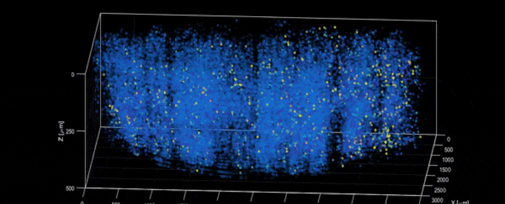
Scientists can see multiple depths in one pulse. This gives them a faster and deeper view of what's going on. Scientists have demonstrated that the technique can track 1,000,000 neurons simultaneously in a mouse brain.
Vaziri says, "There is no reason why we didn’t do it five years ago." It would have been possible if the microscope and laser technology had existed. It was not even thought of."
Scientists hope to use light beads microscopy to observe the interactions between sensory, motor and visual brain regions in mice and other animals.
Understanding and interpreting the neural activity being recorded will take another step, but this new study advances the idea of what is possible with microscopic analysis.
We can see more of the brain and learn more about how it works, whether that's interplay between nerve cells or which brain parts correspond to which emotions.
Vaziri says that light beads microscopy will enable us to examine biological questions in a new way.
Nature Methods published the research.


