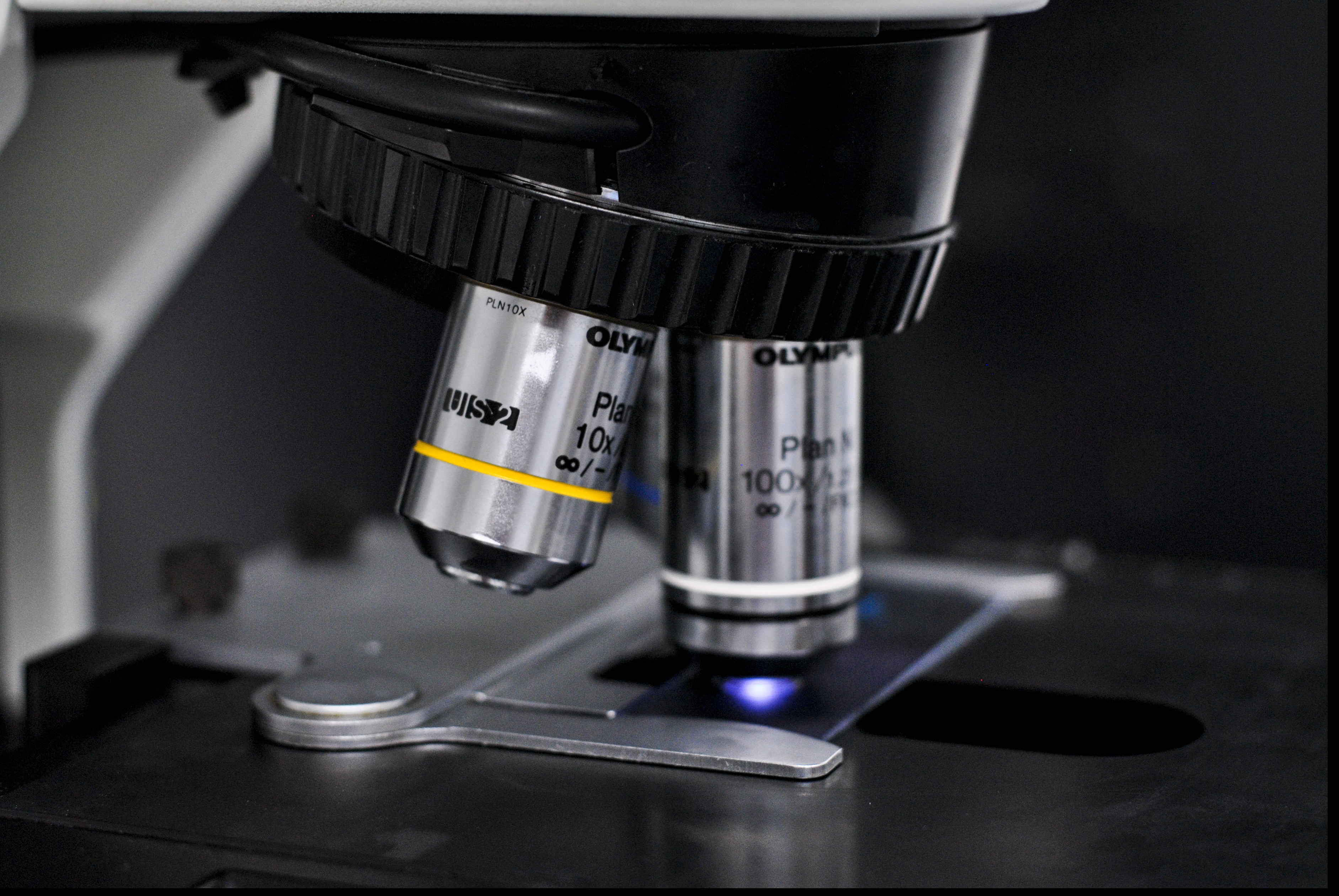
Although there are limits to the information you can get about cells from 2D photos, creating 3D images takes a lot of time and effort. Researchers at UT Southwestern have created a new device that captures multi-angle photos. It can be retrofitted to existing microscopes. Their solution, which requires inserting two rotating mirrors in front a microscope's camera, is 100 times faster than the conversion of images from 2D into 3D.This involves taking hundreds of photographs of the specimen and then uploading them as an image stack to a graphics program. The program performs calculations in order for multiple viewing angles. These two steps can take a lot of time, even with a powerful computer. The team discovered that they could bypass this process by using an optical device.They claim that their method is faster than the traditional approach of using hundreds of frames to create entire 3D image stacks. The technique was discovered while they were de-skewing images taken by two common light-sheet microscopes. They discovered that the projected image would appear to rotate if they applied too much de-skew."This was the eureka! Reto Fiolka is assistant professor at Lyda Hill Department of Bioinformatics, UT Southwestern. We realized that this could go beyond optical de-skewing. The system could also be used with other types of microscopes.The modified microscope allowed the team to image calcium ions that carry signals between nerve cells in a culture plate and examine the circulatory system for a zebrafish embryo. They also quickly captured cancer cells moving and the beating heart of a zebrafish. The optical unit was also used to image additional microscopes such as light-sheet and spinning disc confocal microscopy.


