The Allen Institute for Cell Science, a division of the Allen Institute, put numbers on the internal organization of human cells using hundreds of thousands of high resolution images.
The scientists captured details about the rich variation in cell shape among genetically identical cells. The team described their work in a paper.
The way cells are organized tells us a lot about their behavior and identity. A rigorous way to deal with this kind of organization is missing from the field. We haven't used that information yet.
Biologists can use this study to understand the organization of different kinds of cells. The key organizational principles of the cells the Allen Institute team studies are revealed in the report.
Understanding how cells organize themselves under healthy conditions can help scientists better understand diseases. The image dataset, genetically engineered stem cells, and code that went into this study are all publicly available.
Cells look different even when they are the same type. The same variability that has long plagued the field is an opportunity to study the rules by which a cell is put together. Many others will adopt the same approach because it is generalizable to virtually any cell.
The pear-ness of our cells is computed.
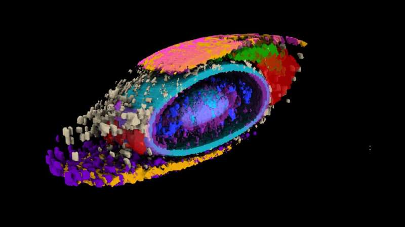
The Allen Institute team first built a collection of stem cells that were genetically engineered to light up different internal structures. Scientists captured high-resolution, 3D images of more than 200,000 different cells with the help of cell lines.
The question is how do our cells organize their interiors.
It turned out that getting to the answer is a lot of work. Imagine setting up your office with hundreds of different pieces of furniture, all of which need to be readily accessed, and many of which need to move freely or interact with one another. Many of the hundreds of pieces of furniture in your office are smaller bags of liquid. It's a nightmare when it comes to interior design.
The scientists wanted to know how the cellular structures fit together. Is structure A always in the same place?
The team was challenged to compare the same structure between different cells. The shapes of the cells under study were different even though they were the same. If one cell was short and blobby and the other was long and pear-shaped, it would be hard for the scientists to compare the positions of the two cells. They put numbers on the pear.
The team developed a shape space that objectively describes the external shape of each stem cell. The shape space has eight different dimensions of shape variation, including height, volume, elongation, andbean-ness. The scientists could look at the organization of cell structures in apples and beans.
Understanding cell shape is important to comprehend how the cells function. "We came up with a framework that allows us to measure a cell's shape, and the moment you do that, you can find cells that are similar shapes, and for those cells you can see how everything is arranged."
The organization is strict.
When they looked at the position of the highlighted structures, they found that all the cells had the same shape. Their internal organization was consistent despite the large variations.
If you look at how thousands of white-collar workers arrange their furniture in a high-rise office building, it's as if every worker puts their desk in the middle of their office and their filing cabinet in the far left corner. If you found an office with a filing cabinet thrown on the floor and papers strewn everywhere, that could tell you something about the state of the office and its occupant.
It's the same thing for cells. Finding deviations from the normal state of affairs could give scientists important information about how cells change when they transition from stationary to mobile, and about what goes wrong in disease. Cells at the edges of colonies of cells and cells that were undergoing division were looked at by the researchers. Scientists were able to find changes in internal organization in the two states.
The executive director of the Allen Institute for Cell Science said that the study brings together everything they've been doing. The metrics to measure and compare different aspects of how cells are organized were built from the ground up. We can now ask questions about cell biology that we could never ask before, and that's what I'm really excited about.
Susanne Rafelski is the author of Integrated intracellular organization and its variations in human i PS cells. www.nature.com/articles/s41586
Journal information: Nature
All Rights Reserved © 2024


