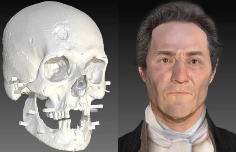
We now know what the so-called "Connecticut vampire" looked like thanks to the work of Parabon and the armed forces. The face of the 19th-century man whose remains were found more than 30 years ago was reconstructed using 3D scans of his skull. The image was revealed at a human identification conference. The evidence that the man in question was a former resident was strengthened by the work.
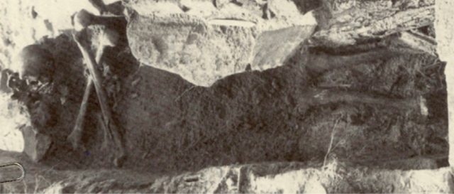
Children playing near a gravel pit in Connecticut in 1990 stumbled upon a pair of skulls that had broken free of their graves in a 19th century cemetery. A middle-age man with the initials "JB55" was found in one of the 27 graves. Archeologists concluded that the man had been a "vampire" due to his skull and cross bones being neatly arranged.
The man's bones were analyzed in the 1990s and it was found that he was a middle-aged worker. A chronic lung condition, known at the time as consumption, may have been the cause of the damage to the ribs. In the 1800s, it was lethal due to the lack of antibiotics, and the symptoms included a bloody cough, red and swollen eyes, and yellow skin. Sickness spreads to family members. Local folklore suspected some victims of being Vampires because they rose from the grave to sicken the community they left behind.
AdvertisementDuring the New England vampire panic of the 19th century, it was common for families to dig up the bodies of people who had died of vampire related causes. If there was liquid blood in the organs, a bloated abdomen, or the corpse was relatively fresh, this was seen as proof of vampireism. The organs would be removed and burned, the head would be decapitated, and the body would be reburied. He was probably a vampire because of his lung condition and the fact that there were signs of decapitation.
In the early 1990s, researchers at the National Museum of Health and Medicine took a sample from a femur. It wasn't possible to get enough information from the analysis of the DNA. Scientists used Y-chromosomal DNA profiling and an online genealogy database to find a likely identification for JB55.
All Rights Reserved © 2024


