The scientists who created the microscope said it could speed up breast cancer treatment.
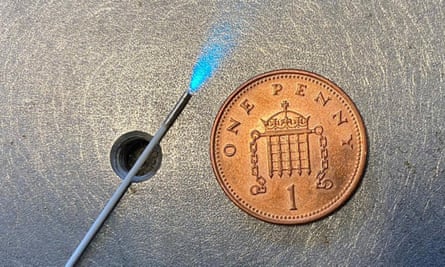
The Imperial College London team has developed a small scope that can be inserted into the body to provide views of tissue and organs.
The team said that the device could produce images from inside the tissue.
The goal is for surgeons to be able to identify cancer cells a hundredth of a millimetre in size at a much faster rate.
The team said that it would help reduce the need for follow up operations.
In breast-conserving surgery, the surgeon removes the cancer while leaving as much normal breast as possible.
Some patients need breast-conserving surgery.
According to the researchers, the device could help cut waiting lists.
They said that using the device would help surgeons identify suspicious tissue very quickly and accurately.
The device is being developed by the Engineering and Physical Sciences Research Council.
The council's director for cross-council programmes, Dr Kedar Pandya, said that by reducing the time it takes to identify cancer cells and improve the accuracy of scans, the endo-microscope developed by Dr Vyas and his team could benefit patients and the National Health Service.
The system is expected to be available for deployment in five years.
The researchers have used their system for preliminary studies on human cancer tissue and are testing its use by surgeons and Pathologists.
All Rights Reserved © 2024


