The university is Kyushu University.
A Kyushu University researcher has developed a new technique for reconstructing plants and animals into 3D models. Over 1,400 models have been made available online.
Stunning pictures of the diverse flora and fauna that encompass our world can be found in nature magazines. From Robert Hooke's sketches of the tiny world in Micrographia to the botanical illustrations in De materia medica, scientists and artists have worked meticulously to draw the true majesty of nature.
The advent of photography has given us more detailed images of animals and plants both big and small, in some cases giving us new information on an animal's structure. Digital libraries grew as technology developed, giving us near unfettered access to valuable data, with methods like computer tomography, orCT, and magnetic resonance scanning becoming powerful tools for studying the internal structure of such creatures.
It's expensive to use magnetic resonance scans and computed toms. Yuichi Kano is an associate professor of Kyushu University's graduate education and research training program in decision science for a sustainable society. "So, we came up with a way to use photogrammetry to create a high-quality 3D image of an animal."
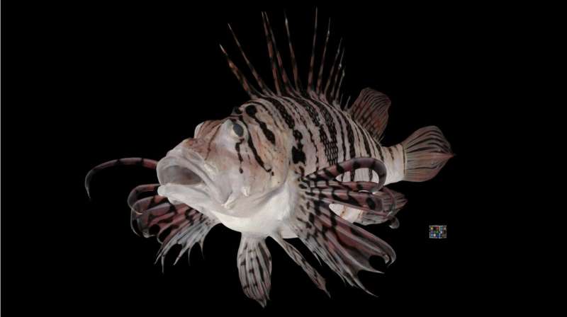
By using photos or other imagery, you can get information and measurement about objects. It can be used to make 3D models similar to what you see on the internet.
The same method was used to make thousands of models of organisms.
We took photos from multiple angles after suspending the sample. Up to 500 of the best photos would be input into the photogrammetry program. It's similar to how the "bullet time" sequence was filmed in the first Matrix movie, except instead of a person, we use an animal.
While he has been working on various organisms, such as insects, plants, and even fungi, he is currently focusing on aquatic animals. Over 1,400 specimen are free to use under the CC BY license.
There are a few limitations in the current methodology, such as difficulty in capturing transparent creatures or making models of exceedingly small organisms, but a few improvements in software and protocols could help.
I hope that this work continues to grow and be used in a variety of fields. It's free to the public so you can use it in education or even use it in a virtual reality machine to explore these organisms up- close. I want to see what people come up with.
More information: Yuichi Kano, Bio-photogrammetry: digitally archiving coloured 3D morphology data of creatures and associated challenges, Research Ideas and Outcomes (2022). DOI: 10.3897/rio.8.e86985 Provided by Kyushu UniversityAll Rights Reserved © 2024


