A new study from North Carolina State University shows a way to study cellular communication using a 3D printer. Learning more about how plant cells communicate with each other is important to understanding more about plant cell functions and could lead to creating better crop varieties and optimal growing environments.
To examine how plant cells acquire and change their identity and function after being bioprinted, the researchers bioprinted cells from the model plant.
The first author of a paper describing the work is a researcher from NC State. Some of the genes being expressed are cell specific. We wanted to know if the cells are alive and doing what they should be doing after being bioprinted.
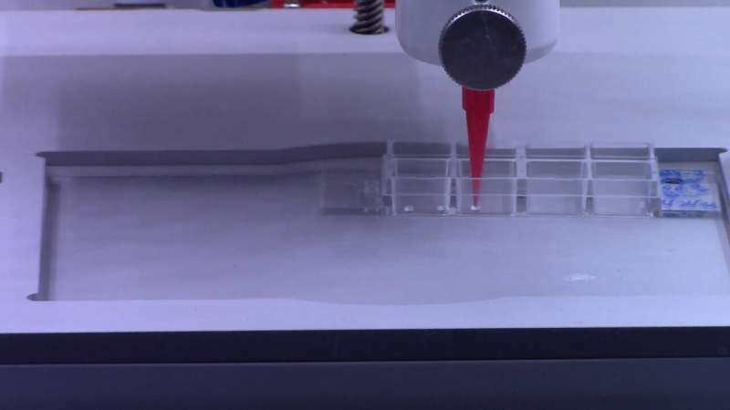
The process of printing plant cells is mechanically similar to printing ink or plastic.
We use living plant cells instead of 3D printing ink or plastic. There are a few differences between the two processes, one of which is the use of an ultraviolet filter to print different bioinks at the same time.
A seaweed-based compound called agarose was added to the live plant cells to make them stronger. Similar to mortar that supports bricks in the wall of a building, agarose provides cells strength and scaffolding.
Ross Sozzani is a professor at NC State and a co-corresponding author of the paper. When you print the bioink, you want it to be liquid but solid when it comes out. Natural environment changes help keep signals and signals in soil.
Microcalli, or small colonies of cells, were formed from more than half of the 3D bioprinted cells.
"We expected good viability on the day the cells were bioprinted, but we had never maintained cells past a few hours after bioprinting, so we had no idea what would happen days later." The 3D printing process doesn't seem to do anything harmful to cells after manually pipetting them.
The pressure of the droplets and the speed at which the droplets are printed are controlled by 3D bioprinting. Control over the architecture of the cells after bioprinting can be achieved using layers or honeycomb shapes.
The cells were bioprinted to see if they could be regenerated or divided. The findings show that the root and shoot cells have different needs.
More than 40% of individual soybean embryonic cells remained viable two weeks after bioprinting.
3D bioprinting can be used to study cellular regeneration in crops.
The researchers studied the cellular identity of the bio printed cells. There are high proliferation rates and a lack of fixed identities in embryology. Similar to animal or human stem cells, these cells can change into different cell types.
Van den Broeck said that bioprinted cells can take on the identity of stem cells by dividing and growing. You bioprint a whole population of cells. We were able to look at the genes expressed by individual cells after 3D bioprinting.
3D bioprinting will allow the researchers to study cellular communication at the single cell level.
The study shows that 3D bioprinting can be used to identify optimal compounds for plant cell viability and communication in a controlled environment.
Science Advances contains the research. The co-corresponding author of the paper is Tim Horn.
More information: Lisa Van den Broeck et al, Establishing a reproducible approach to study cellular functions of plants cells with 3D bioprinting, Science Advances (2022). DOI: 10.1126/sciadv.abp9906. www.science.org/doi/10.1126/sciadv.abp9906 Journal information: Science AdvancesAll Rights Reserved © 2024


