We use cookies to make sure that our website works correctly, as well as some optional cookies to personalize content and advertising, provide social media features and analyse how people use our site. By accepting some or all optional cookies, you agree to the processing of your personal data, including transfer to third parties, in countries that don't offer the same data protection standards as the country where you reside. Clicking on'manage settings' will give you more information about how your data is processed. We have a privacy policy.
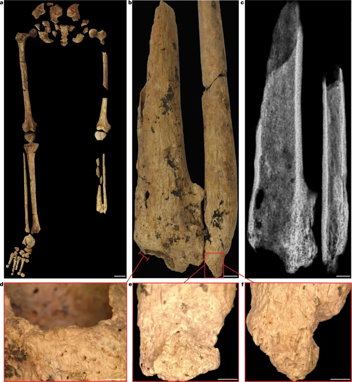
This article is about nature. The emergence of settled agricultural societies around 10,000 years ago gave rise to a host of health problems that had previously been unknown among non-sedentary populations and stimulated the first major innovations in prehistoric medical practices. The oldest known indication of an operation was the removal of a Neolithic farmer's left forearm, which was partially healed. The earliest evidence of a complex medical act is a case of amputation dating to around 7,000 years ago. The remains of a young person from Borneo who had a third of their left leg removed as a child have been found. The person survived the procedure and lived for another 6 years before their remains were buried in a cave in East Kalimantan, Indonesian Borneo, which contains some of the world's earliest dated rock art. The early evidence of a successful limb amputation suggests that some modern humans in tropical Asia had developed advanced medical knowledge and skills long before the Neolithic farming transition. The Sangkulirang–Mangkalihat Peninsula of East Kalimantan is home to an extensive limestone karst landscape that was located close to the easternmost edge of the continental landmass. There is archaeological evidence of prehistoric human occupation in the caves and rock shelters that can be found in this rugged terrain. There is a significant gap in archaeological records of human remains in the region. A large three-chambered limestone cave with preserved rock art is located 2.5 km from and 165 m above the Marang river. A 2 m by 2 m trench was excavated in the central floor of the largest chamber of the cave. The area was excavated to a depth of 1.5 m without reaching bedrock, revealing nine major units and a fully articulated single adult inhumation. Sunda is the continental shelf region that covers the present-day island of Borneo during periods of lowered sea levels. The easternmost edge of Sunda is next to the Sangkulirang–Mang Kalihat Peninsula. There is an area shown in b. The map is from the Shuttle Radar Topography Mission 1 Arc- Second Global by NASA/NGS/USGS. The base maps were created using ArcGIS. A scale bar of 500 km and 10 km. The grave cuts into and modifies SU8 by displaying a strongly defined boundary and distinctive infill. The bottom of the ovate-shaped grave cut did not go into the underlying SU9. The western margins of the burial cut were clearly visible when the western excavation wall was crossed. There were limestone rocks above the head and arm of the person. The burial markers and strong feature boundaries were unique to all other horizontal strata. 2 and 3 have confirmed that the burial was a deliberate human grave. They were laid to rest on their back in an almost north-to-south alignment with the left and right legs flexed. Within a confined space 12 and 13 there is a lot of movement of bones. The cultural materials recovered from the burial include flaked chert artifacts and a 22mm by 17mm lump of red ochre, which was found near the mandible. There is a single adult inhumation. The skull is on the right side of the scale bar. A flexed burial position with the right knee brought to the chest and a complete right foot, and the left knee flexed below the pelvis, with the tibia and fibula underneath. The scale bar is 5 cm Laser scanning and photography were used to remove the burial feature and skeleton. The reassembled skeleton shows 75% bone presence, with all teeth present and intact, and is considered relatively complete in terms of representation of the skeleton and bone. The person is classified as a modern human based on their appearance. The combination of epiphyseal fusion, pubic symphysis, and auricular surface stages, as well as analyses using dental formation techniques, suggest that the person who died was a young adult. The sex is indeterminate due to the cranium and pelvis showing intermediate sex characteristics. When compared with other prehistoric male individuals with a resemblance to pre- Last Glacial skeletons from Asia, the TB1 individual is taller than the mean for most female individuals. The age of the charcoal sample was 31,133 to 30,437 years before the present. An estimate of 31,115 to 30,437 cal. bp was returned by a charcoal sample from the burial feature. The maximum date with an estimate of 31,519 to 31,054 cal. bp is provided by the charcoal recovered from the SU9 area. The SU9 sample was located underneath the burial cut and within a completely different area than the SU8 sample. The age estimate for the burial feature is between 31,519 and 30,437 cal. The boundary between the burial and the underlying SU8 is 31,135 864 cal. bp according to the data. The depth measurement of each sample shows a strong and significant correlation with the age of the sample, and this is confirmed by the radiocarbon dating from overlying stratigraphic units. There is a positive age–depth relationship of these samples that supports an argument for minimal deposit reworking. An age estimate of 25.4 4.3 thousand years old was returned from the analysis of a sample of the left mandibular molar. The analysis in isolation and radiocarbon dating of the remains were unable to be done due to insufficient amounts of radioactive material. Incorporating the electron spin resonance age into the model gives a modelled date of 31,206 to 30,734 years ago, which is 95.4% probability. The oldest intentional primary burial of a modern human is thought to be between 31,000 and 30,000 years old. The left foot was missing from the burial feature. There are 3 and 4 The left fibula shaft fragments were found flexed underneath the left leg. There are four and five. All of the right foot bones were recovered within the grave. The left fibula shaft fragments have amputation surfaces that are covered by bone. There are 4 and 5 supplementary information. This shows that a third of the lower leg was removed through surgery. In cases of modern trauma in which a large metal blade or a mechanical process has been involved, the trauma pattern is not consistent with the descriptions of non- surgical amputation. Non-surgical amputations do not cause clean oblique sectioning and are not clinically recorded to sever the lower limb of both the tibia and fibula, as is the case for Tuberculosis 1 The comminuted and crushing fractures 18 are caused by blunt-force trauma from an accident or an animal attack. The careful treatment of the individual in life after the amputation and in burial is not consistent with someone considered deviant 19 The inferior margin of the fibula has been completely remodelled, which shows that this was not a fatal pathology. The left limb is the most common cause of infections in open wounds. The lack of infections precludes the possibility of an animal attack, such as a crocodile bite, due to the high likelihood of infections from the animal's teeth. There is partial consolidation of the bone between the fibula and tibia. The changes are consistent with late-stage amputations. The small size of the left tibia and fibula is suggestive of a childhood injury as the bones did not continue growing. The heavily restricted use of the left leg is one of the reasons why the left tibia and fibula is thin. The absence of the third leg in the left leg is shown in the picture. The bone is porous because the dead bone was removed. A digital microscope is used to take the images. The scale bars are 5 cm, 5mm, and 2mm. Important implications for our understanding of the evolution of human medico-socio-cultural practices can be found in the surgical amputation of the left lower leg of a person. There isn't much evidence of surgery before written records. Until now, the earliest evidence of advanced medical knowledge, including amputation, was limited to cases from the past. It has been held by many western scholars that healthcare systems and medical procedures of historically known forager societies are rudimentary. Traditional healing practices usually involve a lot of knowledge of plant-based remedies. The development of surgical intervention and treatment of people with illness or injury is thought to have been poorly developed among small-scale communities. It has been assumed that more complex surgeries were beyond the capabilities of the previous and current societies. The removal of body parts is thought to have been limited to phalangeal amputation for symbolic purposes. Historical accounts differ from ancient Roman sources to advances in surgical procedures developed during the past few centuries. The review of the latter 1 and 27 gives details of modern clinical procedures of amputation, demonstrating the level of hygiene, surgical skill, and required apparatus for success, which is synonymous with survival of the person with illness or injury. Within the past 100 years, successful surgical amputation became a medical standard. It was thought that most amputation surgery patients would die at the time of amputation from blood loss and shock or from subsequent infections, since there were no markers of advanced healing left. The amputation of this individual's lower left leg must have been done by a Surgeon who knew how to prevent fatal blood loss and infections. It is necessary to remove the limb for survival. During surgery, the surrounding tissue, including veins, vessels and nerves, were exposed and negotiated in such a way as to allow this individual to live with altered mobility. If the individual was immobile, intensive post-operative nursing and care would have been necessary, such as temperature regulation, regular feeding, bathing, and movement to prevent bed sores. The wound would have been regularly cleaned, dressed, and disinfected in order to prevent infections and provide pain relief. It's not possible to determine if the person had an infectious disease after the surgery, but they probably didn't. Life without a lower limb is thought to be combined with other traumas. A high degree of community care can be assumed to have overcome several of the practical challenges presented by the rugged and mountainous terrain. The discovery of old evidence of deliberate amputation demonstrates the advanced level of medical expertise developed by early modern human foragers. Over a long period of time and through oral traditions of learning, the comprehensive knowledge of the human body is likely to have been developed. It is not known if this was a rare and isolated event in the history of this region or if this particular society had achieved a high degree of skill in this area. The risk of death from trauma and disease has always been with us, and complex medical acts, such as limb amputation, could have been more commonplace in the pre-agricultural past of our species. Poor preservation of pathological bone and preconceptions about theprimitive nature of earlier medico-socio-cultural practices may affect our understanding of this aspect of H. Human colonization of the ancient rainforests of Borneo may have led to the development of medical technology that was unique to this region. The development of new pharmaceuticals may have been stimulated by the rapid rates of wound infections in the tropics. Ground-penetrating radar and electrical resistivity tomography were used in the survey. The data was collected using a Mal X3M with a 500-Mhz antenna and a time window of 62 ns. The GPR data was processed using a suite of filters. The ERT data was collected using a ZZ flash Res-64 with a 1.5 m spacing and Wenner and dipole–dipole array with k values of 20 and a dipole–dipole l value of 5 An on-time of 1.2 s and an off-time of 0.2 s were recorded. The data was output using ZZ RData Check software, inverted in Res2D using the robust scheme and displayed with a colour scale constructed using the Jenks Breaks feature. The features within the deposit were excavated separately from the rest of the deposit. The Homogenous sediments were excavated in units that measured between 1 cm and 5 cm in thickness. A robotic total station was used to record materials and features. All artifacts larger than 19mm in size were plotted in three dimensions and scanned with a laser. The features were sieved using a soft nylon screen, while the rest were sieved using a 1.5mm screen. All artifacts can be traced back to either an excavation unit or a stratigraphical unit. Stone artifacts, shell, faunal remains and macrobotanical remains are among the cultural materials recovered. Human remains and other delicate artifacts were excavated using handheld tools to prevent damage and other delicate artifacts were removed using a leaf trowel. The burial feature was first seen at 0.87 m depth in the western squares. The unit was marked by a very different colour and texture. The western margins of the burial cut were cross-sectioned by the western excavation wall to form a profile. The features of the burial were unique to surrounding and overlying areas. The observations show that the body was not put into natural crevices or deposited through natural processes. The placement of limestone rocks as burial markers further distinguishes the upper surface of the grave. There is a red lump on the left clavicle that is likely to be a dead body. Primary and relatively undisturbed burials are supported by anatomical integrity and articulation of unstable joints. A total of 10 in situ radiocarbon dating samples were dated using AMS 14 C dating at the Direct AMS laboratory in Seattle, USA. The Northern Hemisphere atmospheric curve was used to calibrate the dates. Acid–base–acid protocols were used to pre treat the samples. The samples were put in 6 M HCl at 65 C for 12 minutes and then put back in 6 M HCl at 65 C for 12 more minutes. The base step was repeated multiple times. The pre-treatment was finished with 2 more rinses. The samples received additional base steps for a total of 4 and 5 steps. The sample D-AMS 038338 showed signs of breakdown in base and was treated with a less aggressive acid–base–acid base step and a deionized water rinse. Stable isotope values are not available for these samples. The GARG facility of the Southern Cross University has a US-ESR dating machine. The tooth was cut in half using a diamond saw with a blade of 300 m. To calculate the internal dose rate, the sample was analysed with a laser and a MC-ICPMS NeptuneXT. The Freiberg X-ray irradiation chamber was used to irradiate the fragment. After adjusting for the baseline, the isotropic signals and assessment of the NOCORS contribution were taken into account. The MCDOSE 2.0 software 40 was used to obtain the curve. The age calculations were done with DATA. The age estimates were used to perform the modelling. The analysis included the probability of individual distributions. The model was composed of phases and boundaries in a contiguous pattern. The goal of the modelling was to estimate the age of the boundaries between thegraphical units based on the results of dating. There was no attempt to get rid of identified outliers. It is hard to identify true outliers because we don't know the underlying age depth model and we are using different dating methods. In keeping with the nature of the dating methods and the quality of the results, we have explicitly specified minimum and maximum ages. This is a better method than an outlier analysis as it avoids unnecessary bias and represents a more conservative approach. The uncertainties are relatively small. The identified boundary ages are not sensitive to changes in the model calculation resolution. The age model results were not affected by the changes we made to the model set up. The age of the burial layer was conservatively estimated as the boundary between the base of this layer and the base of SU7 was incorporated. Both completeness and taphonomy were assessed for bone preservation. The Skeletal and Dental completeness and post-depositional processes were assessed. Age-at-death estimation techniques were used to estimate the age of the person. Pubic symphysis and auricular surface degeneration stages were compared. A narrow age estimate of late teenage years to early adulthood can be achieved by different fusion times. Epiphyses that are not fused until later in life, such as the end of the clavicle, were assessed. The age-estimation protocols used dental eruption, wear and formation methods. The maximum length of the bones were estimated using regression equations. The right femur and tibia were considered the most valuable bones because of their relationship to stature. Estimates for pre-Neolithic individuals from southeast Asia are likely to be better if the population is from the Melanesians. The American Black stature estimate standards were used because of the similar proportions to the maximum tibia lengths and 10mm adjustments to the maximum lengths. Estimates for comparative pre-Neolithic hunter-gatherers in southeast Asia have traditionally been estimated from modern Asian populations in the US. The estimates are provided for comparison to other pre-Neolithic humans. An assessment of bone changes was done. Revised standard protocols 54, 55, and 56 were used to record Lesions. The percentage of bone affected by the lesion and the bone type affected were recorded to assess the spatial distribution of the disease. The level of healing, margin definition, presence of necrotic bone, focality, shape changes to the bone, and laterality were recorded to reconstruct the progression and pattern of disease. Lesions were compared against clinical and paleopathological literature to find possible candidates for the disease. To describe the mechanism of injury, force, type and time of trauma, and the degree and complexity of healing, trauma analysis followed previously published protocols. Information on research design can be found in the Nature Research Reporting Summary. This study's data is included in the published article. The Supplementary Table 3 contains all the code used in this study. The Archaeology of Medicine is written by C.A. Roberts. R. Arnott It's the second volume. 1046 was published in the Archaeopress. There is a brief review of the archaeological evidence. The euro is a foreign currency. J. Clinical. Nutrition was published in 2002 A scholar from the internet. The oldest amputation on a human skeleton was in France. The person is called Nat. It was published in the Prec. The paleolithic cave art in Borneo has been published. Nature 556, 257 A scholar from the internet. The Skeletal remains of a modern human were found in Indonesia. The journal PLoS ONE 9 will be published in 2011. A scholar from the internet. The question was posed by O'Connell, J. It's Proc. The National Acad. It's a science The United States of America 115, 8482–490. A scholar from the internet. The handbook of bioarchaeology in Southeast Asia and the Pacific Islands was written by Oxenham and Buckley. The sub-adult burial from Gua Makpan, Alor Island, Indonesia was done in the early Holocene. It's Quat. There is an int. The numbers are 603, 125, and 136 in the years to come. A scholar from the internet. The skull from Niah Cave was excavated in the late 19th century. There is a front It's called ecol. There is an evol. The year was 4,75. A scholar from the internet. The earliest modern human presence in Sumatra was 73,000 years ago. Nature 548, 32–25 A scholar from the internet. The origin of human burial can be traced back to the paleolithic. The view from Qafzeh, Saint-Césaires, Kebara, Amud, and Dederiyeh are not dead issues. It's J.Hum. There is an evol. 37, 27 and 90 were published. A scholar from the internet. The most well-known human burial in Africa is Martinn-Torres. Nature 95– 100 will be published in 2011. A scholar with the search engine. There are immediate and eventual features of healing after amputation. A woman named Ann. 93, 97, 98, and 99 (1929). A scholar from the internet. A review of 127 patients with jet-ski related injuries. It's called orthop. There is traumatol There was a report on the Surg.Res. 104. A scholar from the internet. G P Pennoyer had a traumatic amputation of his thigh. J.Am. is a fictional character. There is a medical field. It's an association The year 1930. A scholar from the internet. BMD and bone geometry are studied in a group of amputees. J. bone miner In Res. 23, there is a description of the situation. A scholar from the internet. A case report on a fractured pars of the axis. There is a person named "Turk." There is a surgical procedure called a neuralsurg. 17th of July 2007, A scholar from the internet. Amputation of limbs. It's cli. It's called orthop. It's relat. Res. 472 was published in 2004. A scholar from the internet. A trauma analysis is done. Is that am? The J.phys. There is anthropol. There were 104 and 139 in 1997. A scholar from the internet. The metabolism of the bones of the tibia in a man was studied. The word orthop is derived from the Greek word for orthop. It was scanned. 33 (Supp.). It was 3– 81 in 1961. A scholar from the internet. Immobilization and bone structure in humans are studied. It's an arch. It's a disease. There are biophysysysysysysysysysysysysysysysysysysysysysysysysysysysy The book is 503, 146–151. A scholar from the internet. There were round-crocodile bites in the country. There is a medical facility in the country of Malawi Med. J. 21, 29 and 31 were published. A scholar from the internet. The skulls were taken from the Copacabana Peninsula in the Titicaca Basin. There is an int. J. paleopathol. There were 9, 20 and 27 in this year. A scholar from the internet. Evidence of trepanation was found in the Yellow River Basin. The word Archeol is derived from the Latin word Archeol. There is anthropol. It's a science The year 2020. A scholar from the internet. The significance of Neanderthal healthcare is discussed in Spikins, P. It's Quat. It's a science The Rev. 217 was published in 2019. A scholar with the search engine. Medical history can be used in medical education. There is a bull in this picture. It's hist. There is a medical field. The year 1947. A scholar from the internet. Burns is the author of the forensic anthropology training manual. Two cases of successful ankle replantation were presented. It's an arch. It's Plast. 47, 182–186 was published in 2020 A scholar from the internet. The traditional knowledge of the Dayak Tribe is used in the use of plants. The biodiversitas 22 will be published in 2011. A scholar from the internet. The Medicinal Plants of Borneo was written by S.P. Gibbons and Teo. Severe disability and paralysis are required in Neolithic Asia. There is anthropol. It's a science A scholar from the internet. The social implications of care provision to seriously disabled individuals were modeled. There is an int. J. paleopathol. The book was 1, 35–42 A scholar from the internet. The Savanna in Borneo was found during the late Pleistocene. It's a science Rep. 9, 6362. A scholar with the search engine. The origin of human burial can be traced back to the paleolithic. The IntCal20 Northern Hemisphere radiocarbon age calibration curve was published in 1997. Radiocarbon 62 was recorded in 2020. A scholar from the internet. The implications of the model for CO 2 radicals in fossil tooth enamel were presented by Grn. It's Quat. There is ageochron 6, 84–97 A scholar from the internet. Grn, Aubert, M., and Moncel analyzed the U-series isotope distribution in a Neanderthal tooth from Payre. There is a person named "geochim" There is a person named "coochim." It's an act. 72, 5279–5289 A scholar with the search engine. There is a detailed protocol for an accurate non-destructive direct dating of a tooth fragment. There are 40geochronometria in the world. A scholar from the internet. There is a new version of the MCDoseE. There is a new program for fitting and evaluating the dose response curve. It's Quat. There is ageochron There were 44, 13 and 22 in this year's edition. A scholar from the internet. The DATA program is used for the calculation of age estimates on teeth. It's Quat. There is ageochron The year was 2009, and it was 4, 231-232 A scholar from the internet. There are standards for data collection from human remains. The Guidelines to the Standards for Recording Human Remains was written by McKinley. It's ch. The Institute of Field Archaeologists published a report in 2004. The Acsdi-Nemeskéri and Suchey-Brooks methods were compared to determine Skeletal age. It's a little bit dull. There is an evol. There were 5 articles in 1990. There is a new method for determining the age at death of adults. Is that am? The J.phys. There is anthropol. In 1985 there wereoldids 69, 15 and 28. A scholar from the internet. There is a laboratory and field manual for juvenile osteology. The excavation, analysis, and interpretation of human remains was written by D.H. Ubelaker. The formation stages of ten permanent teeth were compared. J Dent. Res. 42 was published in 1963. A scholar from the internet. The dental wear scoring technique is used by Scott. Is that am? The J.phys. There is anthropol. 51, 212–117 was published in 1979. A scholar from the internet. The timing of linear hypoplasia on human anterior teeth was brief communication. Is that am? The J.phys. There is anthropol. There were 115, 135, and 140 in 2000. A scholar from the internet. Variation in human formation times. It's J.Hum. There is an evol. 50, 329– 347 A scholar from the internet. Estimation of stature from long bones of white and black people. Is that am? The J.phys. There is anthropol. In1952, 10, 463–528. A scholar from the internet. Secular change in long bone length and proportion in the US from 1800 to 1970. Is that am? The J.phys. There is anthropol. There were 105, 57, and 67 in the year 1999. A scholar from the internet. In forensic approaches to death, disaster and abuse is written by J. and R. The Australian Academic Press published Oxenham's book. Health and Disease in the Prehistoric Pacific Islands was written by H.R. Buckley. The Identification of Pathological Conditions in Human Skeletal Remains was written by D.J. Ortner. The Munsell Color Co. Inc. produced soil color charts. Download references The director of the National Centre for Archaeology in Jakarta authorized the work. The Indonesian State Ministry of Research and Technology is acknowledged by us. The Forrest Foundation supports I.E.D.-H. A fellowship from the Australian Research Council was used to support this research. The research was supported by the Australian Research Council and Southern Cross University. The authors contributed equally were Tim Ryan Maloney, India Ella Dilkes-Hall, Melandri Vlok, Adhi Agus Oktaviana, Pindi Setiawan. The centre for social and cultural research is at the university. Tim Ryan and Maxime Aubert are related. The Australian Research Centre for Human Evolution is located in Nathan. They are: Tim Ryan Maloney, Adam Brumm, and Maxime Aubert. The school of social sciences is located in Western Australia. IndiaElla Dilkes-Hall. The South East Asian Centre is located in the University ofSydney. Melandri V Lok. Pusat Riset Arkeometri is located in Indonesia. Adhiags Oktaviana. The school of humanities, languages and social science is at the university. Adhi Agus Oktaviana. The Bandung Institute of Technology has a faculty of art and design. The person is Pindi Setiawan. The Budaya Kalimantan Timur is located in Indonesia. Muslimin A. R. Effendy, Budi Istiawan, and Falentinus Triwijaya Atmoko are all related. Budaya Berkelanjutan is located in Indonesia. The two men made Geria. The College of Humanities, Arts and Social Sciences is located in South Australia. Ian's name is Ian. The GARG is a research group at Southern Cross University. There are two people, RenaudJoannes-Boyau and Maxime Aubert. The University of Johannesburg has a paleo- research institute. The name of the person is RenaudJoannes-Boyau. T.R.M., I.E.D.-H. and A.A.D.P. wrote the manuscript after excavating the site. The authors contributed to the manuscript. P.S., M.R., A.A.O., F.T.A., I.M.G., M.A.R.E, B.I., and S.A. were involved in facilitating site access, project coordination The US–ESR dating analyses were done by R.J. B. India Ella Dilkes-Hall, Adhi Agus Oktaviana, Pindi Setiawan, Andika Arief Drajat Priyatno, Ian Moffat, and Tim Ryan Maloney are all recipients of correspondence. There are no competing interests declared by the authors. Nature thanks Robin Dennell, Tom Higham and the other anonymous reviewers for their contributions to the peer review of this work. The largest limestone cave in the world is called Tebo. A 2 x 2 m excavation was put in place over a deep deposit in the center of the caves. The area was excavated to a depth of 1.5 m before the project was stopped due to the COVID19 Pandemic. The entire human burial was excavated before the season ended. a, Nine major stratigraphic units (SU) with mostly horizontal bedding and strongly to moderately interpretable boundaries shown across west and north profiles. Sediments were assessed and described using Munsell colour57; compaction (cemented, weakly cemented, moderate, loose to very loose); and sorting grades of well (<0.5 mm), moderate (~1 mm), poor (>1.5 mm), and very poor (>2 mm) particles. Calibrated radiocarbon ages and laboratory codes are presented within respective SUs represented in stratigraphic profile. b, Squares C and D, west wall profile showing burial feature cut that continues into the profile. c, Squares D and A, north wall profile. d, 3D laser scan depicting thorax of TB1 skeleton, with stratigraphic units above the feature (SU5,6,7) depicted on the western wall, and a boundary line of the underlying strata SU9. The figure is shown from left to right. In 32 stages, the burial feature was excavated. There are markers above the skull and arms. There is a skull and a femur under which two bones have been removed. The middle right is the location of the amputation. The burial feature shows grave cut margins. There are healed amputation surfaces of the left and right fibulaes. The white arrows show clear cutting margins. There is a lack of proper veining of the bone tissue. The interosseus is heterogeneously ossified. There was damage to the insect's body. There is a note about the placement of the fibula. There are marks on the left central andlateral incisors. There are two observable defects in the dog. There are two views of the C2 right pars interarticularis. A deep costoclavicular sulcus is associated with the right clavicle. The radiolucent region that extends to the margins of thelysis can be seen on the external bone. The white arrows show that the margins are irregular. The same lysis can't be seen as it can be seen with the femur. It's not visible and is more consistent with an inFracture. The deformity of the original bone shape is caused by (B) being visible. The tooth section and polished Uranium-series analyses show the distribution of uranium in the M3 dental tissues. There are additional notes on the preservation, age-at-death and sex of the Liang Tebo skeleton. There are additional notes on the US–ESR dating analysis. The stature estimates are included in the Supplementary Tables 1–3. You agree to abide by the guidelines for submitting a comment. If you find something that is not in line with our guidelines, please flag it.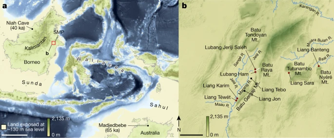

Excavation
Dating
Osteology
Reporting summary
Sunda is the continental shelf region that covers the present-day island of Borneo during periods of lowered sea levels. The easternmost edge of Sunda is next to the Sangkulirang–Mang Kalihat Peninsula. There is an area shown in b. The map is from the Shuttle Radar Topography Mission 1 Arc- Second Global by NASA/NGS/USGS. The base maps were created using ArcGIS. A scale bar of 500 km and 10 km.
All Rights Reserved © 2024


