In the first of its kind, scientists have captured on video all the steps a virus takes as it enters and leaves a living cell.
Scientists used lattice light sheet microscopy, chemical and genetic manipulation and other techniques to achieve the feat.
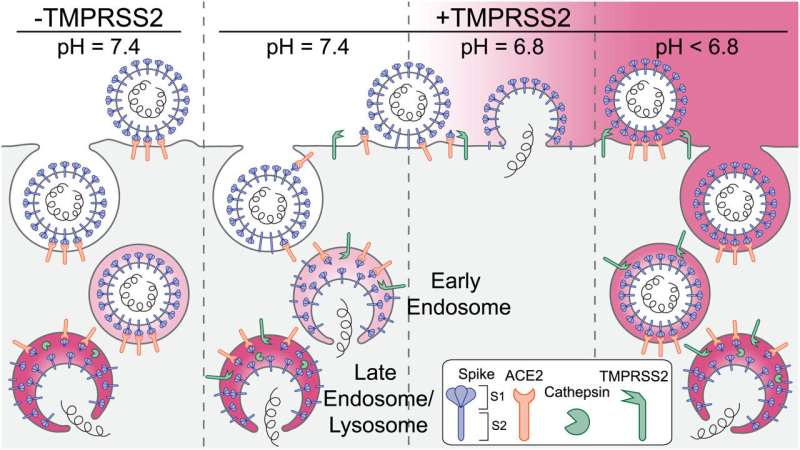
The first part of the video shows a virus that is captured at a cell surface and engulfed by a cellular compartment called an "endosome" The virus injects its genetic material into the cell, which kicks off a cycle of viral infections and replication.
There are many Viruses inside the cell in the second part of the video snapshots are taken every four seconds in the video
The findings published in PNAS provide new insights into the fundamental mechanics of viral infections and could lead to new methods for intervening before the start of COVID-19.
The researchers found that viruses can't release their genomes unless they're bathed in acidic water. The experiments showed that the pH must fall between 6.2 and 6.8, just shy of neutral and on par with bodily fluids. The team's measurements showed that the pH range inside a human nose is similar to the acidity of endosomes.
A professor at Harvard Medical School and Boston Children's Hospital said that measuring the pH of the nostril has never been done before.
The team found that the acidic environment allowed TMPRSS2 to be cut on the cell surface.
The work was done by the labs of the three people who were involved. The first author of the paper is an instructor at the hospital.
More information: Alex J. B. Kreutzberger et al, SARS-CoV-2 requires acidic pH to infect cells, Proceedings of the National Academy of Sciences (2022). DOI: 10.1073/pnas.2209514119 Journal information: Proceedings of the National Academy of SciencesAll Rights Reserved © 2024


