People on the beach are familiar with the pain of a sting from a Jellyfish. How do the stinging cells of jellyfish work? A model for the stinging organelle of the sea anemone has been created by a new research. The study was led by Ahmet Karabulut and was published in Nature Communications. Insights from the work could lead to beneficial applications in medicine.
The Stowers team's model for stinging cell function provides important insight into the architecture and firing mechanism of nematocysts. Both osmotic pressure and elastic energy are involved in piercing and poisoning a target.
In order to understand the structure and operating mechanism of nematocysts, we used a number of different techniques.
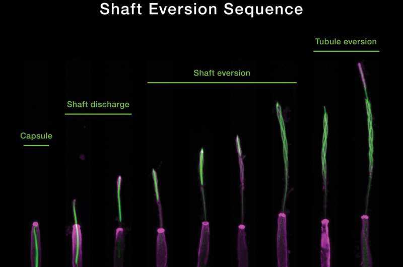
The researchers used state-of-the-art methods to group the process into three separate phases. The nematocyst capsule is the initial target of the first phase. There is a change in osmotic pressure caused by the sudden influx of water and elastic stretching of the capsule The discharge and extension of the thread's shaft substructure is marked by the release of elastic energy through a process called eversion, which is the mechanism where the shaft turns inside out. The tubule begins its own eversion process in the third phase, releasing neurotoxins along the way.
Potential future applications for humans can be found in understanding the stinging mechanism. This could lead to the development of new delivery methods for medicines.
It takes just a few thousandths of a second for the stinging operation to be complete. The firing of the nematocyst is very fast and difficult to capture.
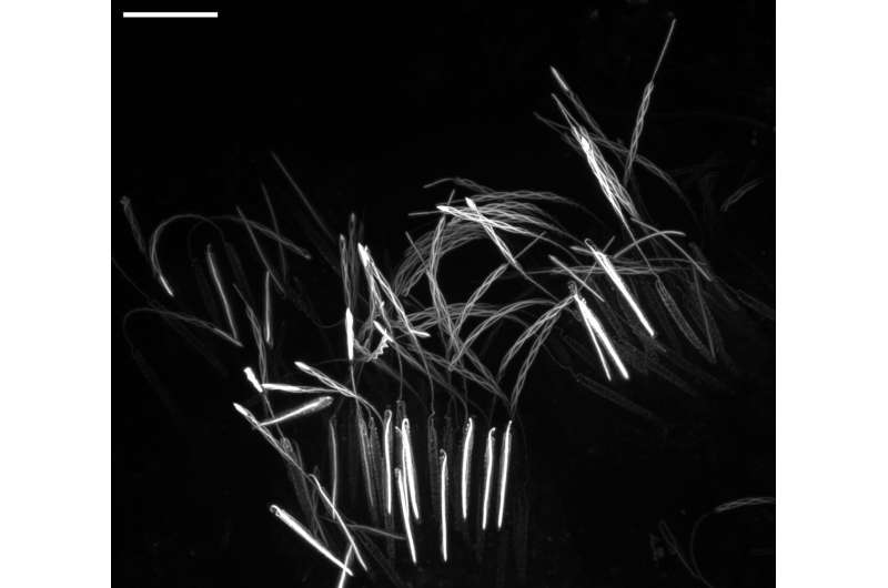
The initial discovery was an accident of curiosity. The fluorescent dye was put into the sea anemone to see what it would look like. He was surprised to have captured multiple nematocysts at different stages of firing after applying a combination of solutions.
I looked under the microscope and saw a picture of a thread discharging. It was a big show. Karabulut said that he realized that nematocysts partially discharged their threads while he used a reagent to fix them.

Karabulut said that he was able to capture images that showed the geometric transformation of the thread. We were able to comprehend the geometric changes of the nematocyst thread.
There are some interesting implications for the design of engineered tiny devices if we are able to understand how nematocyst firing occurs in a sea anemone.
The co-authors are Boris Rubinstein, Melainia McClain, and Sean McKinney.
More information: Ahmet Karabulut et al, The architecture and operating mechanism of a cnidarian stinging organelle, Nature Communications (2022). DOI: 10.1038/s41467-022-31090-0 Journal information: Nature Communications