There is a functional role for a calcium channel in T-cells, according to a study.
The Magerstadt Professor of Pharmacology and co-senior author of the study said that the findings add to a growing body of knowledge about T-cell activation.
"If we have a better understanding of what controls T-cell activation, then we can design approaches to control immune response, both in situations where it is good and where it is bad," said Prakriya, who is also a professor of Medicine.
T-cells are a major component of the body's immune response and are activated by calcium signaling in response to invaders such as foreignbacteria.
The Orai1 calcium channel is used to raise the calcium concentration in T-cells. The hunter-killer functions of T-cells are aided by the production and release of inflammatory mediators.
The calcium channels in neurons are different from Orai1 and play a big role in cellular activation. There are signs that calcium channels may be active in T-cells, but it's not clear if that's true.
In the current study, investigators examined T-cells isolated from both human and mouse tissue and looked for signs of calcium channels and expression of genes. They did not find evidence for the presence of functional calcium channels in human and mouse T-cells. The calcium channel proteins were expressed in the T-cells but not functional. Instead, these transcripts were truncated and degraded.
Prakirya said that no functional voltage-gated calcium signaling was detected in T-cells from human patients with a loss of function.
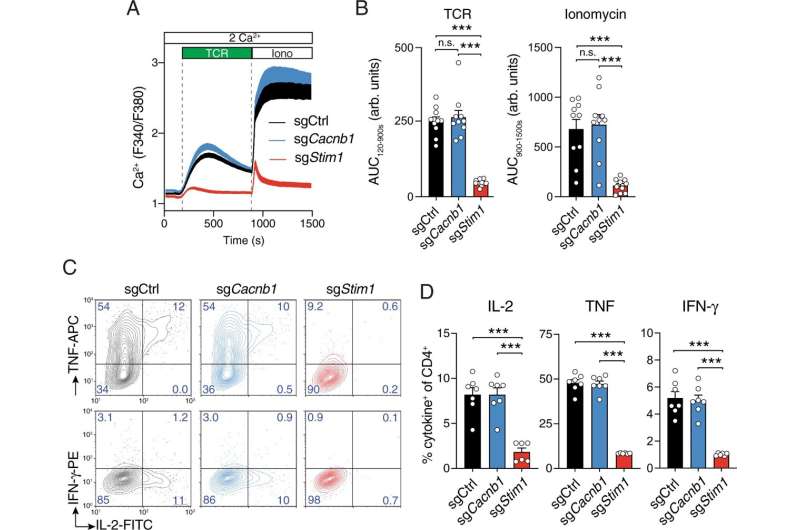
The investigators used gene editing to remove certain calcium channels. The function of a larger calcium channel depends on the function of the alpha and beta subunits.
The investigators found that while the alpha subunit is dispensable, it is necessary for proper activation.
The function of the voltage-gated channel and the function of the T-cell seem to be unrelated to the role played by thebeta subunits. It needs to be better understood.
In the future, better control of T-cell activation could be used to correct an inflammation in the body that can be caused by a variety of diseases.
Understanding the signaling network that controls T-cells is a fertile area for drug discovery as newer approaches to modify T-cell responses can be used for controlling inflammatory and allergic diseases.
More information: Serap Erdogmus et al, Cavβ1 regulates T cell expansion and apoptosis independently of voltage-gated Ca2+ channel function, Nature Communications (2022). DOI: 10.1038/s41467-022-29725-3 Journal information: Nature CommunicationsAll Rights Reserved © 2024


