A Rutgers scientist is part of an international team that has determined the process for incorporating selenium, an essential trace mineral found in soil, water and some foods that increases antioxidant effects in the body.
The most in-depth description of the process by which selenium gets to where it needs to be in cells is included in the research. It's important to note that selenium is encapsulated within selenocysteine. Sec is incorporated into 25 selenoproteins, all of them key to a host of cellular and metabolism processes.
Understanding the workings of these vital mechanisms in such a detailed manner is critical to the development of new medical therapies.
Some of the structures that were revealed in the study are unique to biology.
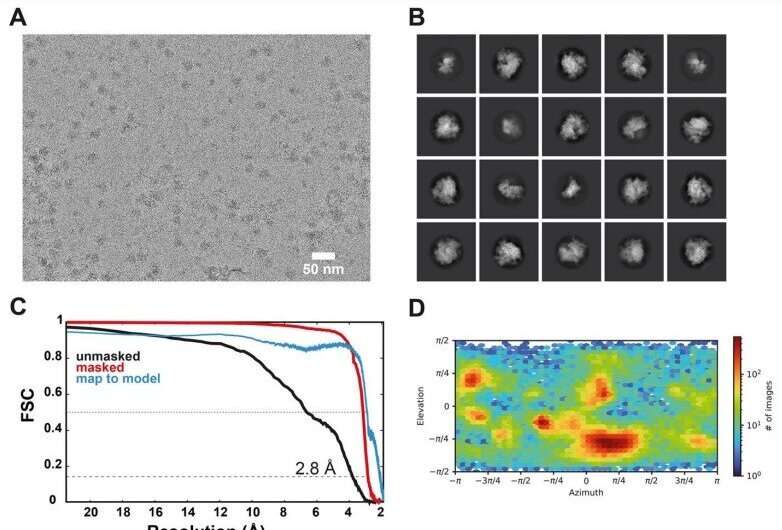
The team was able to see the cell mechanisms using a specialized cryo- electron microscope, which uses beams of electrons rather than light to form three-dimensional images of complex biological formations. The process uses frozen samples of molecular complexes and then applies sophisticated image processing, using today's vast computing power to string together thousands of images to produce three-dimensional cross-sections and even stop- motion animation. Scientists can view representations of the intricate structure of proteins and other biomolecules as they function as cellular "machines."
The cell's intricate machinery requires the inclusion of selenium. Scientists knew which genes were involved in the process. The function of the cell's ribosome is dictated by how these factors work together to complete the cycle. They found that the processes that occur in the human body are not the same as those in other parts of the body.
In order for a biomolecule to function on a biochemical level, it has to be carried to the ribosome via a uniqueprotein factor. In humans, all of this evolved to allow for the inclusion of selenium.
A wide range of vital functions are performed by the selenoproteins once Sec is ensconced in them. The building blocks of DNA are produced by them. They store fat for use in the future. They make cells. The human body's metabolism is controlled by the hormones produced by them. They respond to stress by attacking it with chemicals.
There are a number of diseases and disorders that can arise when the production of selenoproteins is disrupted.
Understanding the mechanism by which Sec is incorporated is a cornerstone of developing new therapies.
More information: Tarek Hilal et al, Structure of the mammalian ribosome as it decodes the selenocysteine UGA codon, Science (2022). DOI: 10.1126/science.abg3875 Journal information: ScienceAll Rights Reserved © 2024


