Detailed knowledge about nature's smallest biological building blocks is needed for the development of new drugs and vaccines. In a completely new way, tiny biological particles can be studied in their natural state with the help of a new microscopy technique.
It takes a lot of time and money to make a medicine or vaccine. It's important to be able to streamline the work by studying how individual proteins interact with each other. The most promising candidates can be found at an earlier stage, thanks to the new microscopy method. The technique can be used to conduct research into how cells communicate with one another by secreting molecule and other biological particles. Our immune response is influenced by these processes.
It is revealing its silhouette.
Since they are the building blocks of everything, biomolecules are important. Researchers need to either mark them with a fluorescent label or attach them to a surface in order to use optical microscopes.
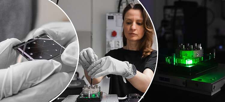
You can't be sure that the surface to which the molecule is attached doesn't affect the molecule's properties. With the aid of our technology, it shows its natural silhouette, which means that we can analyze the molecule just as it is. The new method was developed with researchers in both physics and biology.
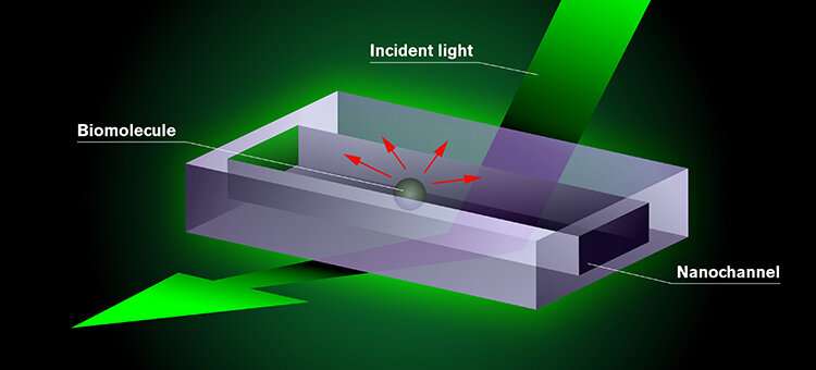
The researchers want to study the molecule or particles that are flushed through the chip with the help of the unique microscopy method. The chip is illuminated with light after a test fluid is added. The molecule can be seen on a screen connected to the microscope when it shows up as a dark shadow because of the interaction between the light and the molecule. It is possible for researchers to see but also determine the mass and size of the biomolecule, something that was not previously possible with a single technique.
The new technique was presented in a journal. The Royal Swedish Academy of Engineering Sciences, which every year lists a number of research projects with the potential to change the world, has paid tribute to the progress made. In this year's Venture Cup competition in Western Sweden, the start-up company Envue Technologies was given the "game-changing" prize.
When you need to study the contents of a sample but don't know what it contains, our method makes the work more efficient.
The researchers are trying to find even smaller particles that are not currently visible.
Langhammer says the aim is to further hone our technique so that it can help to increase our basic understanding of how life works.
The technique is explained.
All Rights Reserved © 2024


