A research team from Columbia University and Carnegie Mellon University have combined two emerging technologies to better view a wide range of biomolecules. Their technique is detailed in Advanced Science.
fluorescent microscopy has its limitations, but it is used to imagemolecules. The tags used in fluorescent microscopy bind to and label the molecule of interest. Researchers can only use 3-4 fluorescent colors in the visible spectrum at a time to label molecule of interest, because these tags emit fluorescent light with a broad range of wavelength.
SRS shows the chemical bonds of biomolecules by capturing their fingerprints. In this way, SRS doesn't need labels to see the different types of biomolecules. A rainbow of dyes can be used to image multiple targets. Many of the important structures found in cells and tissue can't be seen by SRS because of its 300 nanometers limit.
Each type of molecule has its own characteristics. We can see the type of molecule we want by tuning in to the characteristic frequencies of the molecule. Something like changing between the radio stations.
The problem of diffraction limits in a wide range of biological images is addressed by expansion microscopy, a technique that is being developed by the lab. Microscopy expansion transforms biological samples into water-soluble hydrogels. The hydrogels can be expanded to more than 100 times their original volume. Standard techniques can be used to image the expanded samples.
Expansion microscopy surmounts the limitations of SRS just as they were able to surmount the limitations of fluorescence microscopy.
The researchers at Carnegie Mellon and Columbia combined the two techniques to create a Molecule Anchorable Gel-enabled Nanoscale Imaging of Fluorescence. The expansion technique used by Zhao was able to expand samples up to 7.2-fold, allowing them to use a different method to image smaller structures.
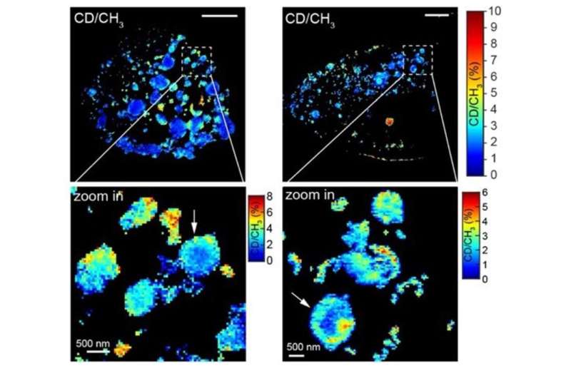
The research team showed in a recently published study that MagNIFIERS could be used for high-resolution metabolic images of the aggregates created in conditions like Huntington's disease. They showed that microscopes could map the location of eight different markers in brain tissue at the same time.
The researchers want to develop a technique that will allow them to understand the pathology of complex diseases, such as cancer and brain disorders.
The additional study co-authors are: Alexsandra Klimas, Brendan Gallagher, Zhangu Cheng, Feifei Fu, Piyumi Wijesekara, and Yupeng Miao.
More information: Lixue Shi et al, Super‐Resolution Vibrational Imaging Using Expansion Stimulated Raman Scattering Microscopy, Advanced Science (2022). DOI: 10.1002/advs.202200315 Journal information: Advanced Science Citation: Researchers magnify hidden biological structures by combining SRS and expansion microscopy (2022, May 18) retrieved 18 May 2022 from https://phys.org/news/2022-05-magnify-hidden-biological-combining-srs.html This document is subject to copyright. Apart from any fair dealing for the purpose of private study or research, no part may be reproduced without the written permission. The content is provided for information purposes only.All Rights Reserved © 2024


