Malaria is caused by single-celled parasites that accumulate in large groups in the salivary glands of mosquitoes. Unless the restriction is lifted by means of appropriate experimental preparation, they can't move. In a set of experiments, researchers at Heidelberg University set the pathogens in motion and analyzed the acquired image data using cutting-edge methods of image processing. The data shows that the pathogens are largely determined by physical principles. Special computer simulations were used to identify the mechanisms behind the rotating movements.
The natural world has a common phenomenon of the collective movement of organisms. The insects and fish move in swarms. Collective movement can also occur at the cellular level, such as when cancer cells migrate from a tumor to form a biofilm. New characteristics that would not otherwise exist in this form can be given rise to by the collaboration of many individuals.
The head of the Institute for Theoretical Physics at the University explains that collectivity creates important processes in physics. He has shown that collective movement can occur in malaria.
The single-celled organisms are injected into the skin through a mosquito bite and then develop in the body. Until now, the collective properties of the cell were not studied. As soon as the mosquito injects the sporozoite into the skin, individual parasites begin to move. Prof. Frischknecht says that this is the critical phase of the infection because it is only successful if the pathogen reaches the blood stream.
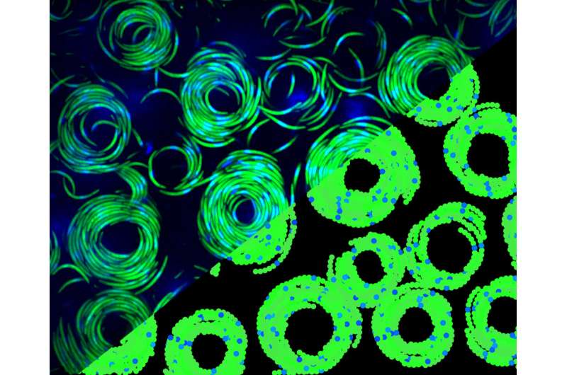
Friedrich Frischknecht and his team discovered that the parasites in salivary glands can be used as a collective. The salivary glands are cut from the mosquito and pressed between two glass plates. The researchers were surprised to find that the crescent-shaped cells were rotating. They are similar to the collective movements ofbacteria or fish, although they differ in that they always move in the same direction. The parasites have a chiral character andfluctuate in size. Prof. Frischknecht believes that the oscillations are due to the fact that they are possible only in the collective of the moving cells.
The data was analyzed to understand them better. Cutting-edge methods of image processing were used by the groups of Karl and Ulrich. They were able to measure the parasites' speed and curvature.
Using computer simulations, it was possible to identify the laws that explain the observations. The interplay of active movement, curved shape of the cell, and chirality in conjunction with mechanical flexibility is enough to explain the sorting and oscillation phenomena in the parasites. The scientists observed that the movement of the individual pathogens resulted in elastic energy being stored in the vortex.
The researchers will investigate how the movement comes about. The structure of sporozoites suggests different possibilities that can be studied. Initial computer simulations show that the right and left-turning parasites quickly separate and build their own systems. Our study shows that the mechanics of the pathogens play an important and heretofore overlooked role.
The research work was done in the framework of the Collaborative Research Center 1129 at the Medical Faculty of the University of Heidelberg. The results of the study were published.
More information: Pintu Patra et al, Collective migration reveals mechanical flexibility of malaria parasites, Nature Physics (2022). DOI: 10.1038/s41567-022-01583-2 Journal information: Nature Physics Citation: Malaria parasites form vortices (2022, May 13) retrieved 13 May 2022 from https://phys.org/news/2022-05-malaria-parasites-vortices.html This document is subject to copyright. Apart from any fair dealing for the purpose of private study or research, no part may be reproduced without the written permission. The content is provided for information purposes only.All Rights Reserved © 2024


