Japan Science and Technology Agency.
Professor Shibata and his team from the University of Tokyo and JEOL were able to observe an atomic magnetic field for the first time in the world. The magnetic-field-free Atomic-ResolutionSTEM was used for the observation. The team was the first to observe the electric field inside atoms. Since the magnetic fields in atoms are very weak compared to electric fields, the technology to observe them has been unexplored. This achievement will rewrite the history of microscope development.
All microscopes have the highest spatial resolution. To achieve ultra-high resolution, we have to place the sample in a strong magnetic field and observe it directly. It has been difficult to observe magnetic materials that are strongly affected by the lens magnetic field. The team was able to develop a completely new structure for the lens in 2019. The team realized atomic observation of magnetic materials, which is not affected by the lens magnetic field. The next goal of the team was to observe the magnetic fields of atoms, which are the origin of magnets.
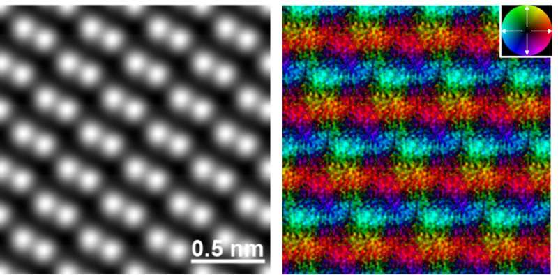
The joint development team took on the challenge of observing the magnetic fields of iron atoms in a hematite crystal by loading MARS with a newly developed high-sensitivity high-speed detector and further using a computer image. The magnetic fields were observed using the Differential Phase Contrast (DPC) method at atomic resolution and a scanning transmission electron microscope. The results showed the origin of magnetism at the atomic level.

The method for observation of atomic magnetic fields was established from the research results. This method is expected to become a new measuring method in the future that will lead the research and development of various magnetic materials and devices such as magnets, steels, magnetic devices, magnetic memory, magnetic semiconductors, spintronics and topological materials.
More information: Yuji Kohno et al, Real-space visualization of intrinsic magnetic fields of an antiferromagnet, Nature (2022). DOI: 10.1038/s41586-021-04254-z Journal information: Nature Provided by Japan Science and Technology Agency Citation: Visualization of the origin of magnetic forces by atomic resolution electron microscopy (2022, February 24) retrieved 24 February 2022 from https://phys.org/news/2022-02-visualization-magnetic-atomic-resolution-electron.html This document is subject to copyright. Apart from any fair dealing for the purpose of private study or research, no part may be reproduced without the written permission. The content is provided for information purposes only.All Rights Reserved © 2024


