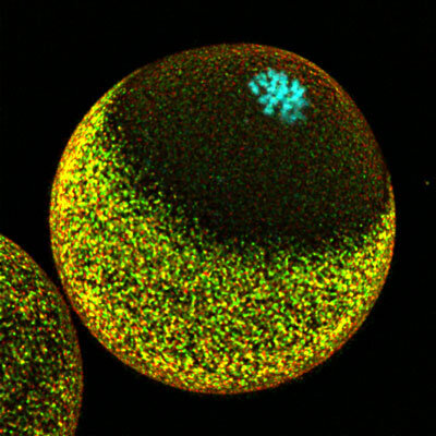
The surface of an unfertilized egg is covered by sperm receptors, except near the maternal chromosomes. The Center for Biosystems Dynamics Research is part of the RIKEN.
The elimination of excess maternal DNA from the egg and the introduction of genomic material from both parents takes place in mice. The finding could help to improve assisted reproductive technologies if the same process occurs in humans.
The end product of meiosis is cells with one copy of each chromosomes. The process of unfertilized ova leaves these cells with both copies of the maternal chromosomes in many animals. One of the extra maternal genome copies is eliminated by meiosis after fertilization.
The presence of maternal DNA alone will cause development to stall if the paternal genome is accidentally ejected. The sorting process has been difficult to untangle. The study of fertilization as a continuous process has been hampered by the lack of robust techniques for high-resolution images of live cells.
The strategy developed by the group allowed them to see the chromosomal dynamics in a mouse egg.
They discovered that the unfertilized egg organizes its internal structure in a way that biases sperm fusion far from the maternal chromosomes. The sperm can be transported to an appropriate fusion site through the formation of structures on the surface of the egg.
The same arrangement of proteins helps to sequester both paternal and maternal DNA. When meiosis restarts, one set of mom's chromosomes will be shed completely, while the other will remain behind to form a complete set with dad's DNA in the newly formed zygote.
The team hopes to find a clinical partner who can help confirm the process in humans. Implications for assisted reproductive technologies could be found if it does. If clinicians aren't aware of where they want to go, a sperm injection in which sperm are directly fused with ova in the laboratory could fail. "In some sperm injections, the sperm may be too close to the mother's chromosomes and the father's chromosomes may be removed," says Mori. The visualization of maternal chromosomes by a non-invasive technique would help to prevent this.
The actin cytoskeleton and the RanGTP keep the paternal and maternal chromosomes apart during fertilization. There is a DOI: 10.083/jcb.202012001.
The Journal of Cell Biology has information.
Successful fertilization requires careful coordination of chromosomes.
The document is copyrighted. Any fair dealing for the purpose of private study or research cannot be reproduced without written permission. The content is not intended to be used for anything other than information purposes.


